Copyright
©2006 Baishideng Publishing Group Co.
World J Gastroenterol. Apr 28, 2006; 12(16): 2620-2621
Published online Apr 28, 2006. doi: 10.3748/wjg.v12.i16.2620
Published online Apr 28, 2006. doi: 10.3748/wjg.v12.i16.2620
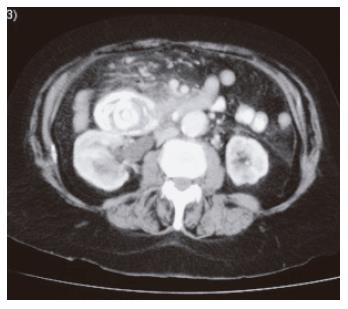
Figure 2 CT abdomen with contrast showing the gall stone in the duodenum with some contrast passing distal to the stone.
- Citation: Masannat YA, Caplin S, Brown T. A rare complication of a common disease: Bouveret syndrome, a case report. World J Gastroenterol 2006; 12(16): 2620-2621
- URL: https://www.wjgnet.com/1007-9327/full/v12/i16/2620.htm
- DOI: https://dx.doi.org/10.3748/wjg.v12.i16.2620


