Copyright
©2006 Baishideng Publishing Group Co.
World J Gastroenterol. Apr 14, 2006; 12(14): 2245-2249
Published online Apr 14, 2006. doi: 10.3748/wjg.v12.i14.2245
Published online Apr 14, 2006. doi: 10.3748/wjg.v12.i14.2245
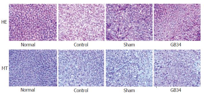
Figure 6 Histological results of liver tissue.
Necrosis and fatty change could be found in the control group (top). GB34 group revealed lower accumulation of extracellular matrix compared to the control group and sham group (bottom) (×400 magnification)
- Citation: Yim YK, Lee H, Hong KE, Kim YI, Lee BR, Kim TH, Yi JY. Hepatoprotective effect of manual acupuncture at acupoint GB34 against CCl4-induced chronic liver damage in rats. World J Gastroenterol 2006; 12(14): 2245-2249
- URL: https://www.wjgnet.com/1007-9327/full/v12/i14/2245.htm
- DOI: https://dx.doi.org/10.3748/wjg.v12.i14.2245


