Copyright
©2006 Baishideng Publishing Group Co.
World J Gastroenterol. Mar 21, 2006; 12(11): 1798-1801
Published online Mar 21, 2006. doi: 10.3748/wjg.v12.i11.1798
Published online Mar 21, 2006. doi: 10.3748/wjg.v12.i11.1798
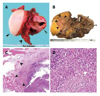
Figure 4 Macroscopically, the cystic mass directly invaded the intercostal space through the diaphragm (A.
Resected crude specimen, black triangle; skin, black arrow head; ribs, white triangle; cystic tumor; B. Cut surface of the specimen, black arrow head; subcutaneous invasion, white triangle; cystic tumor). Microscopically, the bone was destroyed by an invasion of granuloma tissue (C). The granuloma was composed of epithelioid cells with necrotic areas (D).
- Citation: Kawashita Y, Kamohara Y, Furui J, Fujita F, Miyamoto S, Takatsuki M, Abe K, Hayashi T, Ohno Y, Kanematsu T. Destructive granuloma derived from a liver cyst: A case report. World J Gastroenterol 2006; 12(11): 1798-1801
- URL: https://www.wjgnet.com/1007-9327/full/v12/i11/1798.htm
- DOI: https://dx.doi.org/10.3748/wjg.v12.i11.1798


