Copyright
©2006 Baishideng Publishing Group Co.
World J Gastroenterol. Mar 21, 2006; 12(11): 1798-1801
Published online Mar 21, 2006. doi: 10.3748/wjg.v12.i11.1798
Published online Mar 21, 2006. doi: 10.3748/wjg.v12.i11.1798
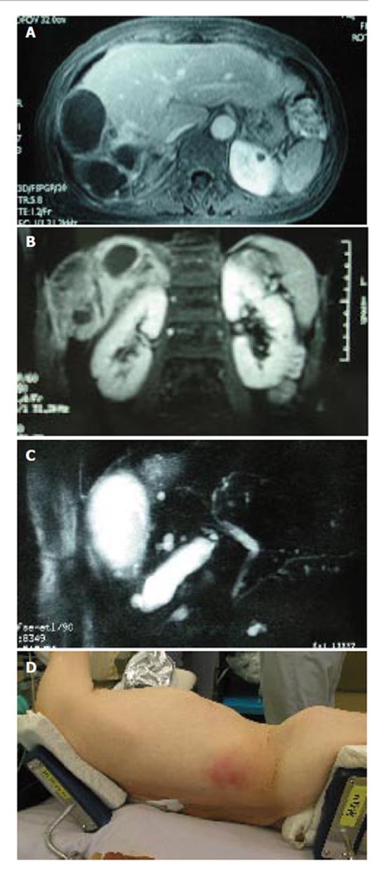
Figure 2 An axial (A) and coronal (B) view of magnetic resonance imaging (MRI) showed a multilocular cyst with a thick wall with a solid component extending into the subcutaneous tissue.
MR cholangio-pancreatography showed no dilatation in the biliary system, and most likely no communication with the liver cyst (C). The skin showed red swelling by the subcutaneous extension of the hepatic cyst (D).
- Citation: Kawashita Y, Kamohara Y, Furui J, Fujita F, Miyamoto S, Takatsuki M, Abe K, Hayashi T, Ohno Y, Kanematsu T. Destructive granuloma derived from a liver cyst: A case report. World J Gastroenterol 2006; 12(11): 1798-1801
- URL: https://www.wjgnet.com/1007-9327/full/v12/i11/1798.htm
- DOI: https://dx.doi.org/10.3748/wjg.v12.i11.1798


