Copyright
©2005 Baishideng Publishing Group Inc.
World J Gastroenterol. Mar 7, 2005; 11(9): 1297-1302
Published online Mar 7, 2005. doi: 10.3748/wjg.v11.i9.1297
Published online Mar 7, 2005. doi: 10.3748/wjg.v11.i9.1297
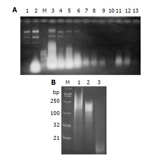
Figure 2 RNAs analysis on 1% agarose gel (A) and 15% non-denaturing polyacrylamide gel (B).
A: RNAs extracted from cells without induction (Lane 1) and with induction with IPTG (Lane 2). The highlight spot represents dsRNAs. dsRNA was purified with CF 11 column as described in materials and methods. Lane 3: RNA extract sample loaded onto CF 11 column; lane 4: flow through; lane 5-10: wash-off with STE containing 18% ethanol (mostly plasmid DNA and single stranded RNA); Lane 11-13: eluate with STE; M: 1 Kb DNA ladder as Marker; B: Digestion analysis of purified dsRNA with RNase A or RNase III. Lane 1: 1 μg undigested purified dsRNA; lane 2: digestion with 0.1 μg RNase A for an hour at 37 °C; lane 3: digestion with 1 U recombinant E. coli RNase III (Ambion) for an hour at 37 °C; M: DNA marker. The length of marker is indicated on the left.
- Citation: Qian ZK, Xuan BQ, Min TS, Xu JF, Li L, Huang WD. Cost-effective method of siRNA preparation and its application to inhibit hepatitis B virus replication in HepG2 cells. World J Gastroenterol 2005; 11(9): 1297-1302
- URL: https://www.wjgnet.com/1007-9327/full/v11/i9/1297.htm
- DOI: https://dx.doi.org/10.3748/wjg.v11.i9.1297


