Copyright
©2005 Baishideng Publishing Group Inc.
World J Gastroenterol. Feb 28, 2005; 11(8): 1155-1160
Published online Feb 28, 2005. doi: 10.3748/wjg.v11.i8.1155
Published online Feb 28, 2005. doi: 10.3748/wjg.v11.i8.1155
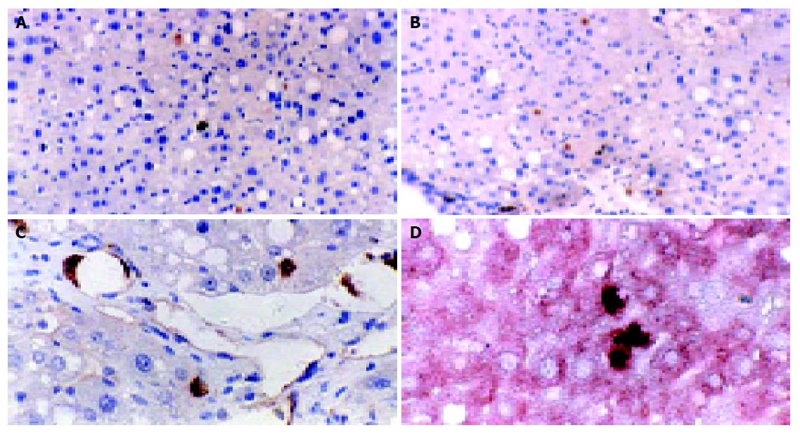
Figure 4 Fate of transplanted BMSCs 4 wk after transplantation.
The differentiation of transplanted BMSCs in liver fibrosis environment. A: Some transplanted stem cells have migrated into parenchyma, exhibiting the morphology of hepatocyte (BMSCs group, 200×); B: Most transplanted cells were distributed at portal region (Guiyuanfang plus BMSC group, 200×); C: Some transplanted stem cells exhibited the morphology of bile epithelium cells or hepatocyte (Guiyuanfang plus BMSC group, 400×); D: Double immunohistochemical staining showed that the BrdU-labeled cells were fully integrated in the liver parenchyma, along with the expression of cytokeratin-18, the liver epithelial specific marker (Guiyuanfang plus BMSCs group, 400×).
- Citation: Wu LM, Li LD, Liu H, Ning KY, Li YK. Effects of Guiyuanfang and autologous transplantation of bone marrow stem cells on rats with liver fibrosis. World J Gastroenterol 2005; 11(8): 1155-1160
- URL: https://www.wjgnet.com/1007-9327/full/v11/i8/1155.htm
- DOI: https://dx.doi.org/10.3748/wjg.v11.i8.1155


