Copyright
©2005 Baishideng Publishing Group Inc.
World J Gastroenterol. Feb 28, 2005; 11(8): 1155-1160
Published online Feb 28, 2005. doi: 10.3748/wjg.v11.i8.1155
Published online Feb 28, 2005. doi: 10.3748/wjg.v11.i8.1155
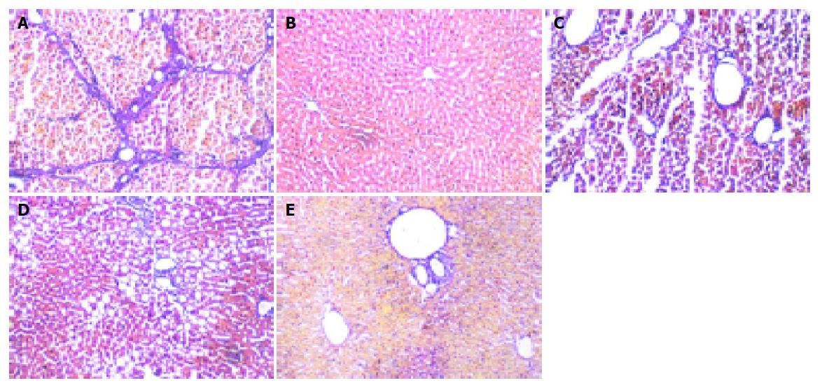
Figure 2 VG staining showing fibrin in liver tissue (100×).
A: In model rats, there was obvious nodular fibrosis with deposition of well-delineated fibrosis septa, extensive collagen deposition, which were continuous and extended throughout each section, with mature collagen fibrils bridging portal regions and vascular structures, including perivenular ballooning degeneration of hepatocytes; B: In control livers, only minimal collagen staining was present; no fibrosis was detected in this group; C: In Guiyuanfang group, collagen deposition decreased; D: In BMSCs group, few collagen deposition. E: The appearance of bridging collagen fibers was prevented almost completely in rats treated with Guiyuanfang plus BMSCs. Only occasional short fibril fragments could be visualized. Most liver tissues were restored to normal organization.
- Citation: Wu LM, Li LD, Liu H, Ning KY, Li YK. Effects of Guiyuanfang and autologous transplantation of bone marrow stem cells on rats with liver fibrosis. World J Gastroenterol 2005; 11(8): 1155-1160
- URL: https://www.wjgnet.com/1007-9327/full/v11/i8/1155.htm
- DOI: https://dx.doi.org/10.3748/wjg.v11.i8.1155


