Copyright
©2005 Baishideng Publishing Group Inc.
World J Gastroenterol. Dec 14, 2005; 11(46): 7277-7283
Published online Dec 14, 2005. doi: 10.3748/wjg.v11.i46.7277
Published online Dec 14, 2005. doi: 10.3748/wjg.v11.i46.7277
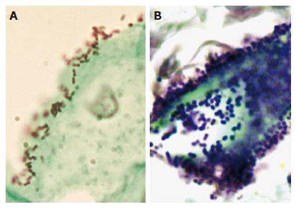
Figure 1 Microscopic examination of bacterial cells in the esophagus.
Esophageal biopsies were fixed in formalin, paraffin-embedded, sectioned, and examined by using Gram-Twort stain. A: In the biopsy from patient #265 with a normal esophagus, Gram-negative cocci and coccobacilli were tightly associated with the surface of squamous epithelial cells. B: In the biopsy from patient #246 with Barrett’s esophagus, Gram-positive cocci were highly concentrated within the lumen of an intestinal-type gland.
- Citation: Pei Z, Yang L, Peek RM, Levine JSM, Pride DT, Blaser MJ. Bacterial biota in reflux esophagitis and Barrett’s esophagus. World J Gastroenterol 2005; 11(46): 7277-7283
- URL: https://www.wjgnet.com/1007-9327/full/v11/i46/7277.htm
- DOI: https://dx.doi.org/10.3748/wjg.v11.i46.7277


