Copyright
©2005 Baishideng Publishing Group Inc.
World J Gastroenterol. Nov 14, 2005; 11(42): 6725-6727
Published online Nov 14, 2005. doi: 10.3748/wjg.v11.i42.6725
Published online Nov 14, 2005. doi: 10.3748/wjg.v11.i42.6725
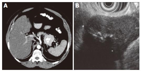
Figure 1 A: Enhanced CT shows a well-circumscribed hypervascular mass of soft tissue density with focal calcification in the tail of pancreas; B: 7.
5 MHz endosonography shows a well-circumscribed hypoechogenic mass (2.8 x 4.9 cm) with focal hyperechogenicity and without signs of infiltration.
- Citation: Goetze O, Banasch M, Junker K, Schmidt WE, Szymanski C. Unicentric Castleman’s disease of the pancreas with massive central calcification. World J Gastroenterol 2005; 11(42): 6725-6727
- URL: https://www.wjgnet.com/1007-9327/full/v11/i42/6725.htm
- DOI: https://dx.doi.org/10.3748/wjg.v11.i42.6725


