Copyright
©2005 Baishideng Publishing Group Inc.
World J Gastroenterol. Oct 28, 2005; 11(40): 6354-6359
Published online Oct 28, 2005. doi: 10.3748/wjg.v11.i40.6354
Published online Oct 28, 2005. doi: 10.3748/wjg.v11.i40.6354
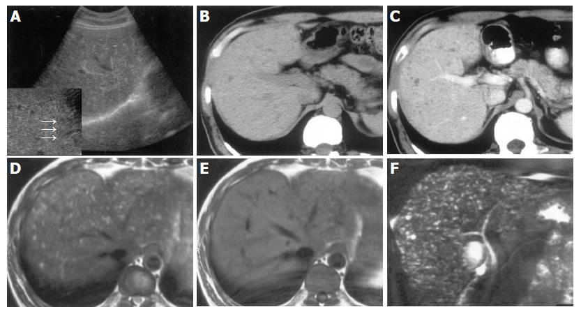
Figure 3 A 44-year-old man with VMCs.
A: B-mode US scan with routine depth showed a vague image of multiple small hyper- and hypoechoic lesions. When magnified by using zoom function, the tiny hypoechoic lesion and the comet-tail echo were clearly seen (arrows); B,C: Multiple tiny hypodense lesions were displayed more conspicuously on enhanced CT (C) than on plain CT (B), and no enhancements were found in the lesions (C); D,E: the lesions.
- Citation: Zheng RQ, Zhang B, Kudo M, Onda H, Inoue T. Imaging findings of biliary hamartomas. World J Gastroenterol 2005; 11(40): 6354-6359
- URL: https://www.wjgnet.com/1007-9327/full/v11/i40/6354.htm
- DOI: https://dx.doi.org/10.3748/wjg.v11.i40.6354


