Copyright
©2005 Baishideng Publishing Group Inc.
World J Gastroenterol. Oct 28, 2005; 11(40): 6354-6359
Published online Oct 28, 2005. doi: 10.3748/wjg.v11.i40.6354
Published online Oct 28, 2005. doi: 10.3748/wjg.v11.i40.6354
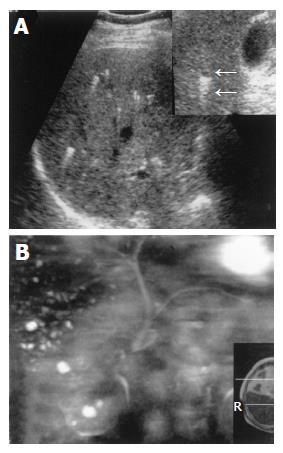
Figure 2 A 72-year-old man with VMCs.
A: On B-mode US, multiple small hyperechoic lesions were shown, while hypo-echoic lesions were not evident on the conventional scanning plan. However, with the use of zoom function, the hypoechoic lesion with comet-tail echo was clearly shown (arrows); B: On MR cholangiopancreatography, multiple small hyper-intensity lesions were revealed except for the demonstration of normal intrahepatic and extrahepatic bile ducts. In addition, several small cysts in the right kidney were also shown.
- Citation: Zheng RQ, Zhang B, Kudo M, Onda H, Inoue T. Imaging findings of biliary hamartomas. World J Gastroenterol 2005; 11(40): 6354-6359
- URL: https://www.wjgnet.com/1007-9327/full/v11/i40/6354.htm
- DOI: https://dx.doi.org/10.3748/wjg.v11.i40.6354


