Copyright
©The Author(s) 2005.
World J Gastroenterol. Oct 14, 2005; 11(38): 6066-6068
Published online Oct 14, 2005. doi: 10.3748/wjg.v11.i38.6066
Published online Oct 14, 2005. doi: 10.3748/wjg.v11.i38.6066
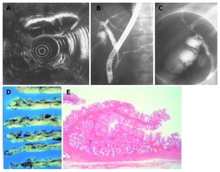
Figure 1 A: Endoscopic ultrasonography of the gallbladder showing multiple thin septa in the neck portion.
B: Endoscopic retrograde cholangiopancreatography showing anomalous pancreaticobiliary ductal union. The common bile duct was not dilated. C: Endoscopic retrograde cholangiopancreatography showing multiple faint septation in the neck of the gallbladder. D:Longitudinal resected gallbladder shows multiple thin septa appearance at the neck portion. E: Microscopic findings of the resected gallbladder demonstrate the septa lined by normal typical columnar epithelium containing normal muscular layer (H&E stain, original magnification ×40).
- Citation: Yamamoto T, Matsumoto J, Hashiguchi S, Yamaguchi A, Sakoda K, Taki C. Multiseptate gallbladder with anomalous pancreaticobiliary ductal union: A case report. World J Gastroenterol 2005; 11(38): 6066-6068
- URL: https://www.wjgnet.com/1007-9327/full/v11/i38/6066.htm
- DOI: https://dx.doi.org/10.3748/wjg.v11.i38.6066


