Copyright
©2005 Baishideng Publishing Group Inc.
World J Gastroenterol. Sep 21, 2005; 11(35): 5577-5581
Published online Sep 21, 2005. doi: 10.3748/wjg.v11.i35.5577
Published online Sep 21, 2005. doi: 10.3748/wjg.v11.i35.5577
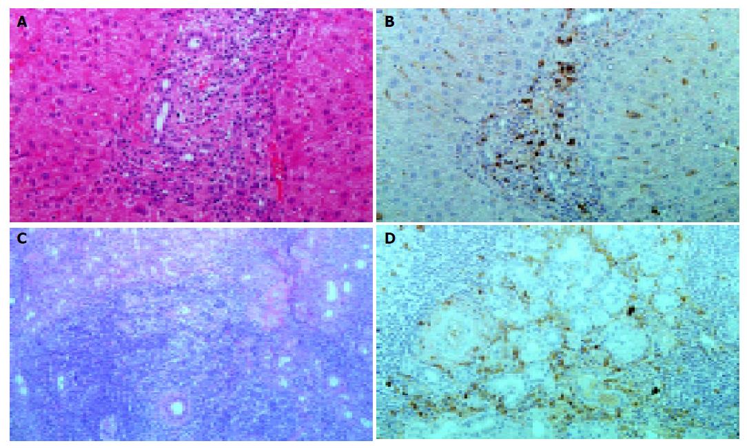
Figure 5 Histopathological examination of the liver (A and B) and salivary gland (C and D) specimens obtained by needle biopsy.
Note the infiltrated lymphocytes and IgG4-positive plasma cells in the portal area, around the ducts of the salivary gland (A and C: hematoxylin and eosin staining, B and D: immunostaining of IgG4).
- Citation: Taguchi M, Aridome G, Abe S, Kume K, Tashiro M, Yamamoto M, Kihara Y, Nakamura H, Otsuki M. Autoimmune pancreatitis with IgG4-positive plasma cell infiltration in salivary glands and biliary tract. World J Gastroenterol 2005; 11(35): 5577-5581
- URL: https://www.wjgnet.com/1007-9327/full/v11/i35/5577.htm
- DOI: https://dx.doi.org/10.3748/wjg.v11.i35.5577


