Copyright
©The Author(s) 2005.
World J Gastroenterol. Sep 7, 2005; 11(33): 5174-5179
Published online Sep 7, 2005. doi: 10.3748/wjg.v11.i33.5174
Published online Sep 7, 2005. doi: 10.3748/wjg.v11.i33.5174
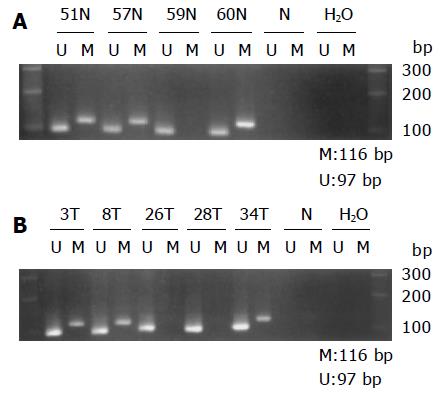
Figure 3 Gel electrophoresis picture demonstrating aberrant methylation in E-cad.
Primer sets used for simple amplification are designed as unmethylated (U), methylated (M), unmodified DNA (N). Water is used as a negative control (H2O). Molecular weight marker is 100-bp DNA ladder. A: Methylation in non-cancerous epithelium. Samples 51, 57, and 60 N are methylated. Sample 59 N is unmethylated; B: Methylation in cancerous lesion. Samples 3, 8, and 34 T are methylated. Samples 26 and 28 T are unmethylated.
- Citation: Liu YC, Shen CY, Wu HS, Chan DC, Chen CJ, Yu JC, Yu CP, Harn HJ, Shyu RY, Shih YL, Hsieh CB, Hsu HM. Helicobacter pylori infection in relation to E-cadherin gene promoter polymorphism and hypermethylation in sporadic gastric carcinomas. World J Gastroenterol 2005; 11(33): 5174-5179
- URL: https://www.wjgnet.com/1007-9327/full/v11/i33/5174.htm
- DOI: https://dx.doi.org/10.3748/wjg.v11.i33.5174


