
















©The Author(s) 2005.
World J Gastroenterol. Aug 28, 2005; 11(32): 5075-5078
Published online Aug 28, 2005. doi: 10.3748/wjg.v11.i32.5075
Published online Aug 28, 2005. doi: 10.3748/wjg.v11.i32.5075
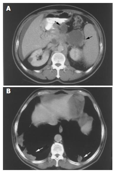
Figure 2 Enhanced CT scan shows multiple, non-enhanced, pancreatic cystic mass (straight and curved arrows) with soft tissue part (A).
multiple, hypodense, alveolar patches at bilateral basal lung parenchyma (straight and curved arrows) (B).
- Citation: Leung TK, Lee CM, Wang FC, Chen HC, Wang HJ. Difficulty with diagnosis of malignant pancreatic neoplasms coexisting with chronic pancreatitis. World J Gastroenterol 2005; 11(32): 5075-5078
- URL: https://www.wjgnet.com/1007-9327/full/v11/i32/5075.htm
- DOI: https://dx.doi.org/10.3748/wjg.v11.i32.5075