Copyright
©The Author(s) 2005.
World J Gastroenterol. Aug 14, 2005; 11(30): 4655-4660
Published online Aug 14, 2005. doi: 10.3748/wjg.v11.i30.4655
Published online Aug 14, 2005. doi: 10.3748/wjg.v11.i30.4655
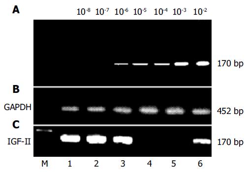
Figure 1 Amplification of IGF-II genomes from human hepatoma tissues or circulating blood samples of patients with hepatocellular carcinoma.
IGF-II mRNAs were synthesized according to IGF-II cDNA with random hexamers and moloney murine leukemia virus reverse-transcriptase, and detected with different primer pairs by nested PCR (170 bp). The positive fragments of IGF-II genome were found distinctly in hepatoma tissues or in peripheral blood of patients with hepatocellular carcinoma. A: the sensitive limitation of our detection system (2 ng/L), using total RNA with 10-2-10-8 fold dilution and then amplified by nested PCR; B: the amplified fragments (452 bp) of glyceraldehyde-3-phosphate dehydrogenase genome from liver tissues or peripheral blood as controls; C: the amplification of IGF-II genomes in liver tissues (No. 1-4) or circulating blood (No. 5-6). No. 1-2, the positively amplified fragments of IGF-II mRNA from cancerous tissues of patients with hepatocellular carcinoma; No. 3, the positively amplified fragments of IGF-II mRNA from para-cancerous tissue of patients with hepatocellular carcinoma; No. 4, no positively amplified fragment from non-cancerous tissue of patients with hepatocellular carcinoma; No. 5, no positively amplified fragment from circulating blood of patients with liver cirrhosis, and No. 6, the positively amplified fragment from peripheral blood of patients with hepatocellular carcinoma. GAPDH: glyceraldehyde-3-phosphate dehydrogenase. M: DNA molecular weight marker.
- Citation: Dong ZZ, Yao DF, Yao DB, Wu XH, Wu W, Qiu LW, Jiang DR, Zhu JH, Meng XY. Expression and alteration of insulin-like growth factor II-messenger RNA in hepatoma tissues and peripheral blood of patients with hepatocellular carcinoma. World J Gastroenterol 2005; 11(30): 4655-4660
- URL: https://www.wjgnet.com/1007-9327/full/v11/i30/4655.htm
- DOI: https://dx.doi.org/10.3748/wjg.v11.i30.4655


