Copyright
©2005 Baishideng Publishing Group Co.
World J Gastroenterol. Jan 21, 2005; 11(3): 457-459
Published online Jan 21, 2005. doi: 10.3748/wjg.v11.i3.457
Published online Jan 21, 2005. doi: 10.3748/wjg.v11.i3.457
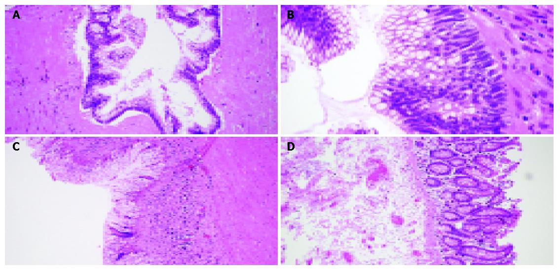
Figure 3 Histological findings.
A and B: Appendix, mucin-filled lumen lined with atypical mucin-producing columnar epithelium and no infiltration in muscular wall, H&E, ×40, ×60; C: Submucosa loaded with chronic inflammatory cells, H&E, ×20; D: Normal colonic mucosa in the cecum, and thickened submucosa, H&E, ×20.
- Citation: Lakatos PL, Gyori G, Halasz J, Fuszek P, Papp J, Jaray B, Lukovich P, Lakatos L. Mucocele of the appendix: An unusual cause of lower abdominal pain in a patient with ulcerative colitis-. A case report and review of literature. World J Gastroenterol 2005; 11(3): 457-459
- URL: https://www.wjgnet.com/1007-9327/full/v11/i3/457.htm
- DOI: https://dx.doi.org/10.3748/wjg.v11.i3.457


