Copyright
©2005 Baishideng Publishing Group Co.
World J Gastroenterol. Jan 21, 2005; 11(3): 377-381
Published online Jan 21, 2005. doi: 10.3748/wjg.v11.i3.377
Published online Jan 21, 2005. doi: 10.3748/wjg.v11.i3.377
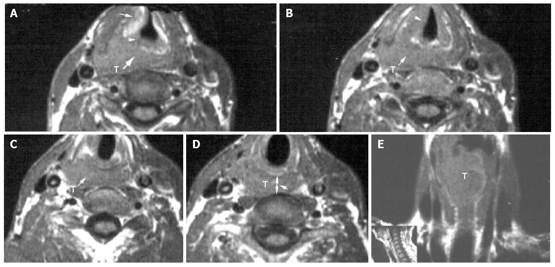
Figure 2 T1-weighted image of a 72 year-old man with pyriform sinus carcinoma invading the esophageal inlet.
A: Axial T1-weighted image of a right-sided pyriform sinus tumor mass (T) invading the right false cord, the laryngeal ventricle, the right paraglottic space (arrow), the right aryepiglottic fold (arrowhead) and the postcricoid region (heavy arrow); B: Axial T1-weighted image of the tumor mass (T) involving the right true vocal cord (arrowhead) and extending to the postcricoid region (arrow); C: Axial T1-weighted image 5 mm above the esophageal inlet level of a pyriform sinus tumor mass (T) invading the postcricoid region (arrow); D: Axial T1-weighted image of the neoplastic esophageal inlet involvement (arrow). T = tumor mass, arrowhead points to the distance between the posterior aspect of the cricoid cartilage and the anterior aspect of the vertebra (d-CV=1.32); E: Conoral T1-weighted image of a tumor mass (T) arising from the right piriform sinus with extension to the postcricoid region and the anteriorlateral wall of the esophagus, including esophageal inlet and cervical esophagus.
- Citation: Chen B, Yin SK, Zhuang QX, Cheng YS. CT and MR imaging for detecting neoplastic invasion of esophageal inlet. World J Gastroenterol 2005; 11(3): 377-381
- URL: https://www.wjgnet.com/1007-9327/full/v11/i3/377.htm
- DOI: https://dx.doi.org/10.3748/wjg.v11.i3.377


