Published online Jun 7, 2005. doi: 10.3748/wjg.v11.i21.3329
Revised: August 28, 2004
Accepted: December 1, 2004
Published online: June 7, 2005
- Citation: Kircali B, Saricam T, Ozakyol A, Vardareli E. Endoscopic biopsy: Duodenal ulcer penetrating into liver. World J Gastroenterol 2005; 11(21): 3329-3329
- URL: https://www.wjgnet.com/1007-9327/full/v11/i21/3329.htm
- DOI: https://dx.doi.org/10.3748/wjg.v11.i21.3329
We have read with interest the recent report by E Kayacetin and S Kayacetin of ‘’Gastric ulcer penetrating to liver diagnosed by endoscopic biopsy[1] since we diagnosed the duodenal ulcer which penetrated into liver similarly. This is a rather unusual case because of the fifth case in the literature and responding to medical therapy.
Eighty-five-year-old female was admitted with 1-mo history of periumblical pain, weight loss, 1-wk vomitting provoked with food and no history of GI bleeding. On physical examination, there was periumblical tenderness.
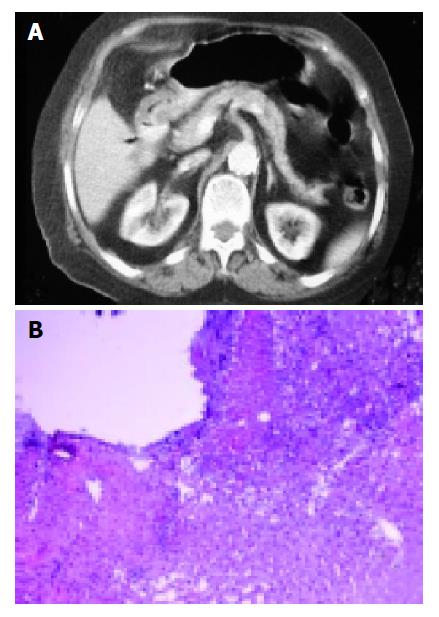
Laboratory evaluation showed Hb: 11.5 gr/dL, WBC: 11000/mm3 MCV: 92 fl, Plt: 415000/mm3, normal liver chemistries, and tumor markers. Grade IV esophagitis and discolerated atypical giant ulcer that stemmed from anterior wall of bulbus duodeni were detected in gastroduodenoscopy. However, it could not be passed through the second part of duodenum due to the presence of edema. Endoscopic biopsy of the ulcer revealed muscular layer without mucosa and exudate on the surface of the liver fragments and macro-microvesicular degeneration, pseudoaciner transformation and perisinusoidal fibrosis in the liver tissue. Furthermore, CT showed fibrofatty on the level of hepatic flexura adjacent to bulbus in the liver (Figure 1).
After 10-d decompression of stomach and intravenous PPI treatment, diminished edema was seen and was easily passed through the second part of the duodenum in the second EGD.
To summarize, in significant part of the patients with abdominal pain without bleeding, penetration peptic ulcer into the liver is diagnosed falsely negative. For this reason, suspicion criteria of penetration of peptic ulcer into the liver should be reviewed and also be developed reliable, cheep diagnostic methods.
Science Editor Guo SY Language Editor Elsevier HK
| 1. | Kayacetin E, Kayacetin S. Gastric ulcer penetrating to liver diagnosed by endoscopic biopsy. World J Gastroenterol. 2004;10:1838-1840. [PubMed] [Cited in This Article: ] |


