Copyright
©2005 Baishideng Publishing Group Inc.
World J Gastroenterol. May 28, 2005; 11(20): 3020-3026
Published online May 28, 2005. doi: 10.3748/wjg.v11.i20.3020
Published online May 28, 2005. doi: 10.3748/wjg.v11.i20.3020
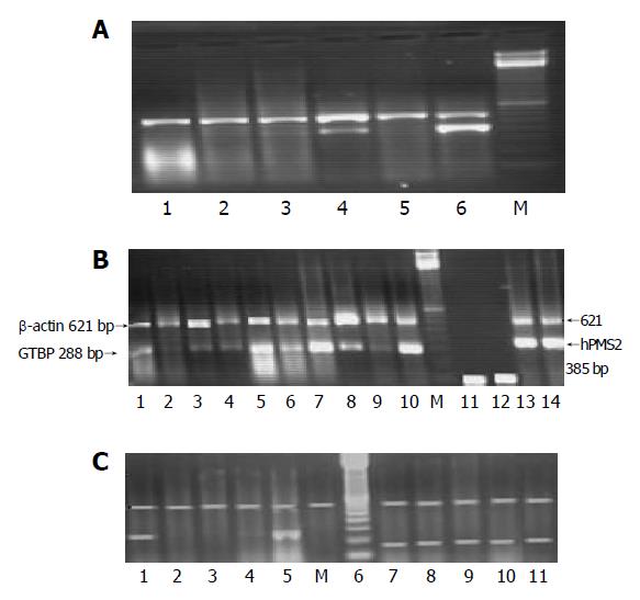
Figure 1 A: PCR amplification for MMR genes.
A: Multiplex RT-PCR of hMSH2 gene in some HCC cases. M: 100-bp marker; lanes 1-6: hMSH2 co-amplified with β-actin in HCC cases; lanes 1-3 and 5: hMSH2 reduction; lane 4: 50 reduced expression of hMSH2; lane 6: hMSH2 co-amplified with β-actin gene in normal PBL as a control. The accepted band of β-actin was at 621 and 492 bp for hMSH2. B: Multiplex RT-PCR of GTBP and hPMS2 genes in some HCC cases. M: 100-bp marker; lanes 1-10: GTBP co-amplified with β-actin in HCC cases; lanes 1 and 6: no reduction; lanes 2, 3, 4, 8 and 9: different expression levels of GTBP gene; lane 5: GTBP co-amplified with β-actin gene in normal PBL as a control; lane 10: overexpression of GTBP; lane 11: GTBP co-amplified with β-actin without NA as reagent control; lane 12: hPMS2 co-amplified with β-actin without NA as reagent control; lanes 13 and 14: overexpression of hPMS2. C: Multiplex RT-PCR of hMLH1 and hPMS1 genes in HCC cases. Lane 1: hMLH1 co-amplified with β-actin gene in normal PBL as a control; lanes 2, 3 and 6: reduced expression of hMLH1 gene; lane 5: overexpression of hMLH1. M: 100-bp marker; lanes 7-10: hPMS1 co-amplified with β-actin gene in HCC cases with no changes on the expression level.
-
Citation: Zekri ARN, Sabry GM, Bahnassy AA, Shalaby KA, Abdel-Wahabh SA, Zakaria S. Mismatch repair genes (
hMLH1 ,hPMS1 ,hPMS2 ,GTBP/hMSH6 ,hMSH2 ) in the pathogenesis of hepatocellular carcinoma. World J Gastroenterol 2005; 11(20): 3020-3026 - URL: https://www.wjgnet.com/1007-9327/full/v11/i20/3020.htm
- DOI: https://dx.doi.org/10.3748/wjg.v11.i20.3020


