Copyright
©2005 Baishideng Publishing Group Inc.
World J Gastroenterol. Apr 21, 2005; 11(15): 2218-2223
Published online Apr 21, 2005. doi: 10.3748/wjg.v11.i15.2218
Published online Apr 21, 2005. doi: 10.3748/wjg.v11.i15.2218
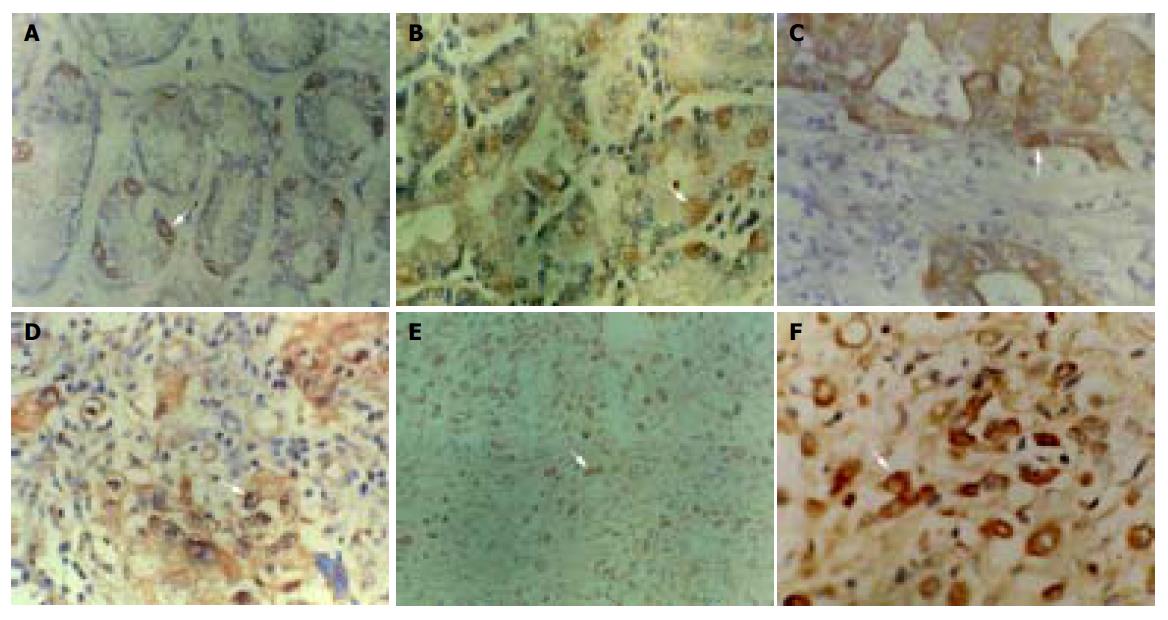
Figure 1 Immunohistochemical staining of P16 protein expression.
Arrow shows positive cell. A: Normal gastric mucosa SP ×400; B: dysplastic gastric mucosa SP ×400; C: well-differentiated adenocarcinoma SP ×400; D: poorly-differentiated adenocarcinoma SP ×400; E: undifferentiated carcinoma SP ×200; F: undifferentiated carcinoma SP ×400.
-
Citation: He XS, Rong YH, Su Q, Luo Q, He DM, Li YL, Chen Y. Expression of
p16 gene and Rb protein in gastric carcinoma and their clinicopathological significance. World J Gastroenterol 2005; 11(15): 2218-2223 - URL: https://www.wjgnet.com/1007-9327/full/v11/i15/2218.htm
- DOI: https://dx.doi.org/10.3748/wjg.v11.i15.2218


