Copyright
©2005 Baishideng Publishing Group Inc.
World J Gastroenterol. Apr 14, 2005; 11(14): 2142-2147
Published online Apr 14, 2005. doi: 10.3748/wjg.v11.i14.2142
Published online Apr 14, 2005. doi: 10.3748/wjg.v11.i14.2142
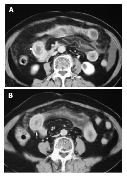
Figure 5 Axial contrast enhanced CT showed rim-calcified ectopic gallstone in the proximal ileum (A).
CT section located 3 cm caudal to (A) reveal segmental edematous wall thickening of small intestine (arrow) proximal to transition zone, which indicated transient ischemic change secondary to bowel obstruction (B) (case 2).
- Citation: Yu CY, Lin CC, Shyu RY, Hsieh CB, Wu HS, Tyan YS, Hwang JI, Liou CH, Chang WC, Chen CY. Value of CT in the diagnosis and management of gallstone ileus. World J Gastroenterol 2005; 11(14): 2142-2147
- URL: https://www.wjgnet.com/1007-9327/full/v11/i14/2142.htm
- DOI: https://dx.doi.org/10.3748/wjg.v11.i14.2142


