Copyright
©2005 Baishideng Publishing Group Inc.
World J Gastroenterol. Apr 14, 2005; 11(14): 2101-2108
Published online Apr 14, 2005. doi: 10.3748/wjg.v11.i14.2101
Published online Apr 14, 2005. doi: 10.3748/wjg.v11.i14.2101
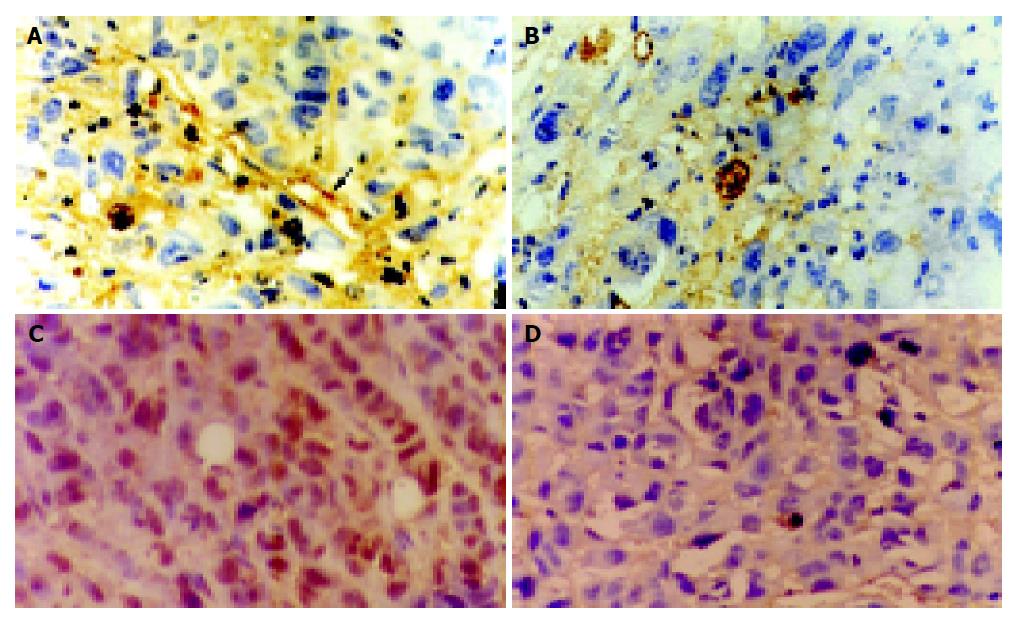
Figure 2 Results of immuno-histochemistry assay of CD34 expression in Pc-3.
A: Plenty of stained and densely arranged micro-vessels in tumor field (arrow); B: After medication the tumor cells loosely arranged in tumor field with scanty of micro-vessels stained or no micro-vessels (SABC ×400); PCNA expression in Pc-3. C: In control site, tumor cells were densely arranged with poly nuclei stained in deep brown yellow; D: In each of the dosed sites loosely arranged tumor cells merely with few nuclei stained in light brown yellow color (SABC ×400).
- Citation: Liu L, Feng GS, Gao H, Tong GS, Wang Y, Gao W, Huang Y, Li C. Chromic-P32 phosphate treatment of implanted pancreatic carcinoma: Mechanism involved. World J Gastroenterol 2005; 11(14): 2101-2108
- URL: https://www.wjgnet.com/1007-9327/full/v11/i14/2101.htm
- DOI: https://dx.doi.org/10.3748/wjg.v11.i14.2101


