Copyright
©2005 Baishideng Publishing Group Inc.
World J Gastroenterol. Apr 7, 2005; 11(13): 2045-2047
Published online Apr 7, 2005. doi: 10.3748/wjg.v11.i13.2045
Published online Apr 7, 2005. doi: 10.3748/wjg.v11.i13.2045
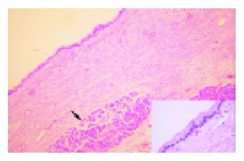
Figure 3 Cyst wall lined by single layered cuboidal to columnar epithelium, rimmed by fibrocollagenous stroma.
Pancreatic acini are seen (arrow). Inset shows high magnification of the cyst epithelium, with no evidence of dysplasia (hematoxylin and eosin).
- Citation: Goh BK, Tan YM, Tan PH, Ooi LL. Mucinous nonneoplastic cyst of the pancreas: A truly novel pathological entity? World J Gastroenterol 2005; 11(13): 2045-2047
- URL: https://www.wjgnet.com/1007-9327/full/v11/i13/2045.htm
- DOI: https://dx.doi.org/10.3748/wjg.v11.i13.2045


