Copyright
©2005 Baishideng Publishing Group Inc.
World J Gastroenterol. Apr 7, 2005; 11(13): 2041-2044
Published online Apr 7, 2005. doi: 10.3748/wjg.v11.i13.2041
Published online Apr 7, 2005. doi: 10.3748/wjg.v11.i13.2041
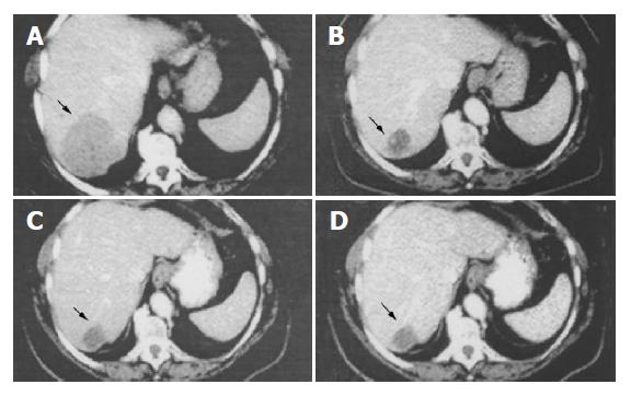
Figure 1 Abdominal CT scan obtained before initiation of SR-LAN therapy (A) showing the liver metastasis (black arrow) at the VI hepatic segment (diameter 70×60 mm).
CT scan obtained after 3 (B), 6 (C) and 18 (D) mo of SR-LAN demonstrates a significant reduction in the size of liver metastasis.
- Citation: Bondanelli M, Ambrosio MR, Zatelli MC, Cavazzini L, Al Jandali Rifa’y L, degli Uberti EC. Regression of liver metastases of occult carcinoid tumor with slow release Lanreotide therapy. World J Gastroenterol 2005; 11(13): 2041-2044
- URL: https://www.wjgnet.com/1007-9327/full/v11/i13/2041.htm
- DOI: https://dx.doi.org/10.3748/wjg.v11.i13.2041


