Copyright
©The Author(s) 2004.
World J Gastroenterol. Dec 1, 2004; 10(23): 3409-3413
Published online Dec 1, 2004. doi: 10.3748/wjg.v10.i23.3409
Published online Dec 1, 2004. doi: 10.3748/wjg.v10.i23.3409
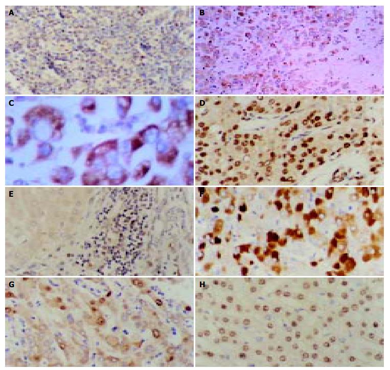
Figure 3 Results of immunohistochemistry.
A: Positive-control of LMP1 from the tonsil tissue. The cytoplasm of lymphocytes showed positive signals. × 400, B: Immunohistochemical staining with anti-LMP1 antibody of an HCC specimen. The positive signals were localized in the cytoplasm and membrane of neoplasm cells. × 100, C: Immunohistochemical staining with anti-LMP1 antibody of an HCC specimen. The positive signals were localized in the cytoplasm and membrane of neoplasm cells. × 400, D: Immunohistochemical staining with anti-LMP1 antibody of an HCC specimen. The positive signals were localized in the nuclei of neoplasm cells. × 400, E: Immunohistochemical staining with anti-LMP1 antibody of an HCC specimen. The positive signals were localized in the interstitial lymphocytes but no clear positive signals were present in the adjacent neoplasm cells. × 400, F: Immunohistochemical staining with anti-HBsAg antibody of an HCC specimen. The positive signals were localized in the cytoplasm of neoplasm cells. × 400, G: Immunohistochemical staining with anti-HBcAg antibody of an HCC specimen. The positive signals were localized in the cytoplasm and nuclei of neoplasm cells. × 400, H: Immunohistochemical staining with anti-HCV antibody of an HCC specimen. The positive signals were localized in the nuclei of neoplasm cells. × 400.
- Citation: Li W, Wu BA, Zeng YM, Chen GC, Li XX, Chen JT, Guo YW, Li MH, Zeng Y. Epstein-Barr virus in hepatocellular carcinogenesis. World J Gastroenterol 2004; 10(23): 3409-3413
- URL: https://www.wjgnet.com/1007-9327/full/v10/i23/3409.htm
- DOI: https://dx.doi.org/10.3748/wjg.v10.i23.3409


