
















©The Author(s) 2004.
World J Gastroenterol. Nov 15, 2004; 10(22): 3318-3321
Published online Nov 15, 2004. doi: 10.3748/wjg.v10.i22.3318
Published online Nov 15, 2004. doi: 10.3748/wjg.v10.i22.3318
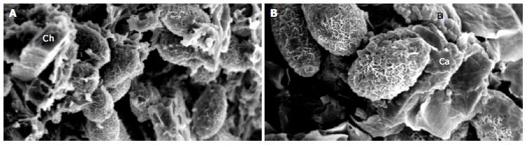
Figure 3 SEM micrographs of gallstones showing Opisthorchis eggs with typical musk-melon-eggshell surface in the nidi of the stones.
Several crystalline structures consistent with calcium (Ca), bilirubin derivatives (Bi) and cholesterol (Ch) could be noted (A, B). Higher magnification with highlighting calcium bilirubinate deposition on Opisthorchis eggshell and mucus is shown in Figure 3B.
- Citation: Sripa B, Kanla P, Sinawat P, Haswell-Elkins MR. Opisthorchiasis-associated biliary stones: Light and scanning electron microscopic study. World J Gastroenterol 2004; 10(22): 3318-3321
- URL: https://www.wjgnet.com/1007-9327/full/v10/i22/3318.htm
- DOI: https://dx.doi.org/10.3748/wjg.v10.i22.3318