Copyright
©The Author(s) 2004.
World J Gastroenterol. Nov 1, 2004; 10(21): 3141-3145
Published online Nov 1, 2004. doi: 10.3748/wjg.v10.i21.3141
Published online Nov 1, 2004. doi: 10.3748/wjg.v10.i21.3141
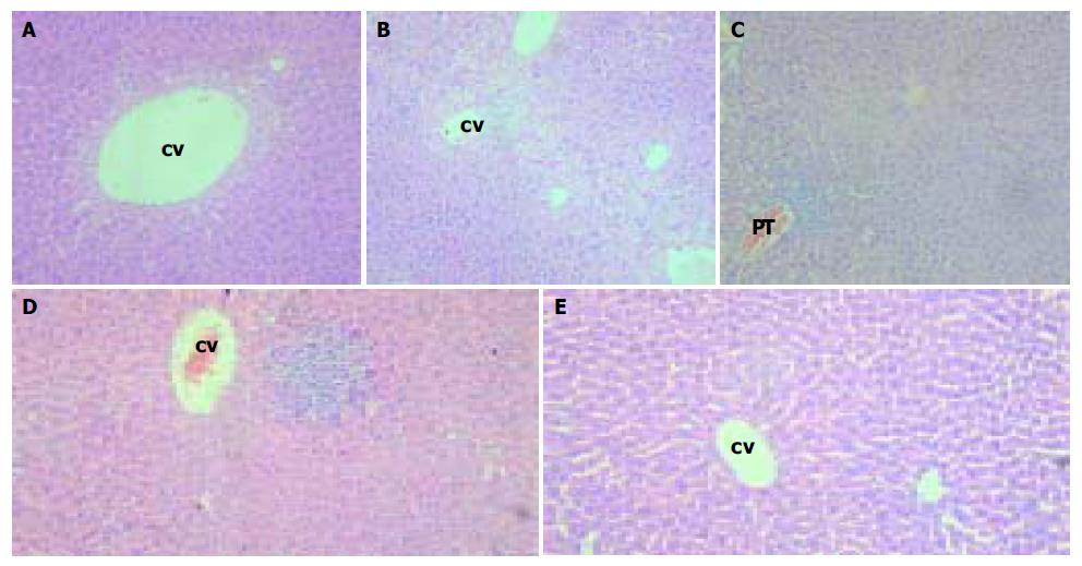
Figure 4 Analysis of pathological changes in the liver of transgenic mice by HE staining.
Magnification × 100. A: Mild swollen hepatocytes around the central veins (CV) in the centrilobular region. B: “Ballooning degeneration” around the central veins or between the two central veins in the liver. C: Predominantly lymphocytic cell infiltrates in portal tract area and hepatic lobules. D: Focal necrosis and predominantly lymphocytic cell infiltrates in hepatic lobule. E: Liver of normal mice.
-
Citation: Ge JH, Zhang LZ, Li JX, Liu H, Liu HM, He J, Yao YC, Yang YJ, Yu HY, Hu YP. Replication and gene expression of mutant hepatitis B virus in a transgenic mouse containing the complete viral genome with mutant
s gene. World J Gastroenterol 2004; 10(21): 3141-3145 - URL: https://www.wjgnet.com/1007-9327/full/v10/i21/3141.htm
- DOI: https://dx.doi.org/10.3748/wjg.v10.i21.3141


