Copyright
©The Author(s) 2003.
World J Gastroenterol. Sep 15, 2003; 9(9): 2025-2029
Published online Sep 15, 2003. doi: 10.3748/wjg.v9.i9.2025
Published online Sep 15, 2003. doi: 10.3748/wjg.v9.i9.2025
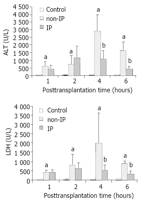
Figure 1 Changes in serum ALT (A) and LDH (B) levels.
The serum ALT and LDH levels were significantly elevated in non-IP groups compared with sham-operated group (aP < 0.01, non-IP group vs sham-operated group). After IP, the increase was significantly reduced at 4 and 6 h points after reperfusion (bP < 0.05, IP group vs non-IP group).
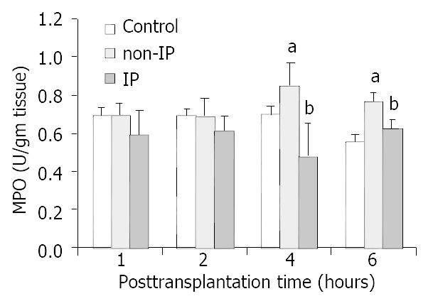
Figure 2 Changes of PMNs accumulation in grafted liver.
PMNs accumulation was assessed by investigating the levels of MPO. The increase in non-IP groups was significantly re-duced in IP groups, especially at 4 and 6 h points after reperfusion (aP < 0.05, non-IP group vs sham-operated group. bP < 0.05, IP group vs non-IP group).
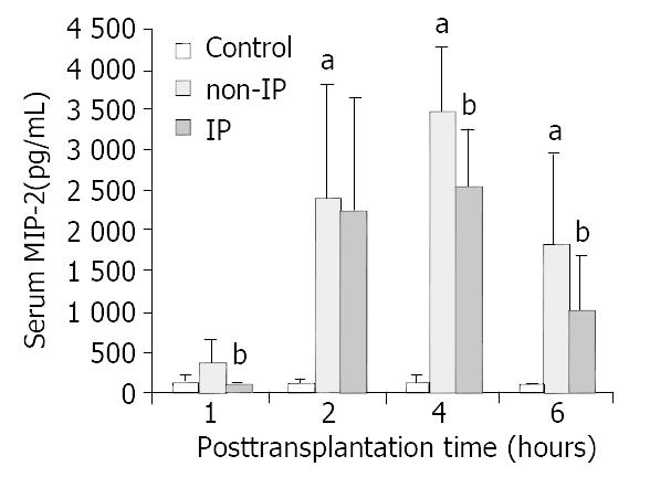
Figure 3 Changes in serum MIP-2 levels.
MIP-2 was signifi-cantly increased in non-IP group compared with sham-oper-ated group (aP < 0.01, non-IP group vs sham-operated group). After IP, the increase was significantly reduced at 1, 4 and 6 h after reperfusion (bP < 0.05, IP group vs non-IP group).
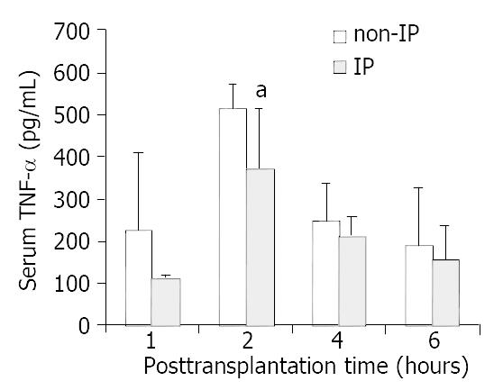
Figure 4 Changes in serum TNF-α levels.
The levels of serum TNF-α were decreased to an extent in each time point respectively in IP group compared with non-IP group, especially at 2 h after reperfusion. The decrease was significant (aP < 0.05, IP group vs non-IP group).
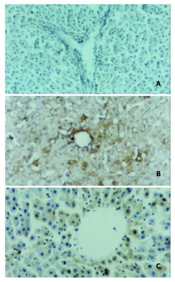
Figure 5 Intragraft MIP-2 mRNA expression.
In situ hybrid-ization was performed at 4 h point after reperfusion in all three groups. MIP-2 mRNA was not detectable in sham oper-ated group (A), and remarkably up-regulated in non-IP group (B), and after IP, the expression was detectable at a lower level (C). MIP-2 mRNA was mostly expressed in hepatocytes in grafted livers.
- Citation: Jiang Y, Gu XP, Qiu YD, Sun XM, Chen LL, Zhang LH, Ding YT. Ischemic preconditioning decreases C-X-C chemokine expression and neutrophil accumulation early after liver transplantation in rats. World J Gastroenterol 2003; 9(9): 2025-2029
- URL: https://www.wjgnet.com/1007-9327/full/v9/i9/2025.htm
- DOI: https://dx.doi.org/10.3748/wjg.v9.i9.2025


