Copyright
©The Author(s) 2003.
World J Gastroenterol. May 15, 2003; 9(5): 915-920
Published online May 15, 2003. doi: 10.3748/wjg.v9.i5.915
Published online May 15, 2003. doi: 10.3748/wjg.v9.i5.915
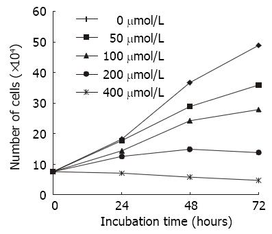
Figure 1 Effects of various concentration of nimesulide on the proliferation of SGC-7901 cells for different time periods analysing by cell counts (n = 3).
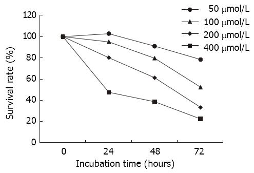
Figure 2 Effects of nimesulide on SGC-7901 cells analyzing the cell survival using the MTT assay.
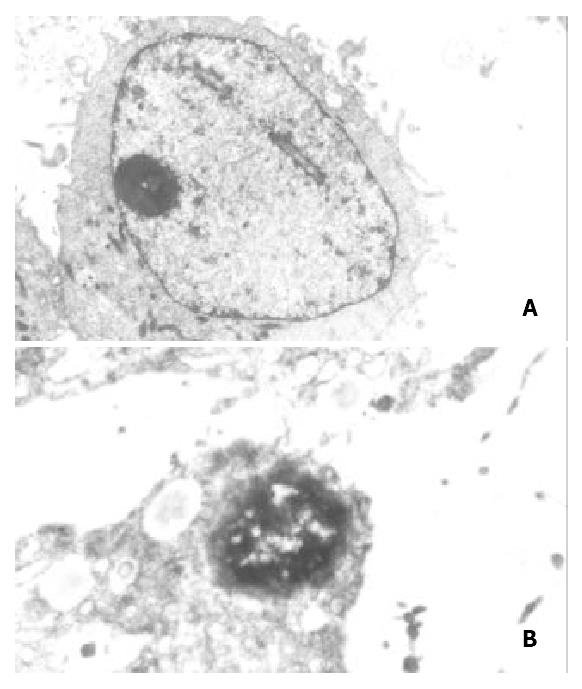
Figure 3 (A) control group: Microvilli of SGC-7901 cells were enrichment, nucleus was large and round, chromatin was disperse, nucleolus was distinct.
TEM × 6000. (B) Treated with nimesulide at the concentration of 400 μmol/L for 72 h: Disap-pearance of microvilli, margination of nuclear chromatin, the organelle swelled up and physallization. TEM × 8000.
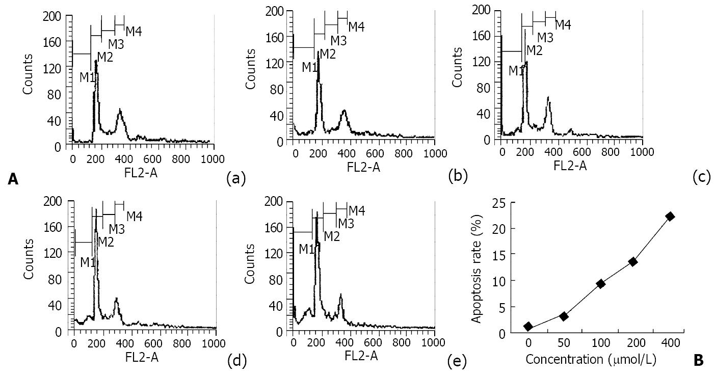
Figure 4 The results of flow cytometry analysis of SGC-7901 cells treated with nimesulide for 72 h and their DNA content was determined by flow cytometry.
A: DNA histogram: (a) control, (b) 50 μmol/L nimesulide, (c) 100 μmol/L nimesulide, (d) 200 μmol/L nimesulide, (e) 400 μmol/L nimesulide. B: The apoptosis percentage of nimesulde-treated SGC-7901 cells.
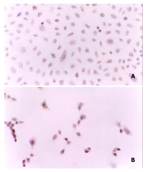
Figure 5 (A) Positive staing of P27kip1 protein was mainly in cytoplasm of SGC-7901 cells before treated with nimesulide.
(B) Strong positive staing of P27kip1 protein was in cytoplasm and in nuclei of of SGC-7901 cells after treated with nimesulide at the concentration of 400 μmol/L. SP × 200.
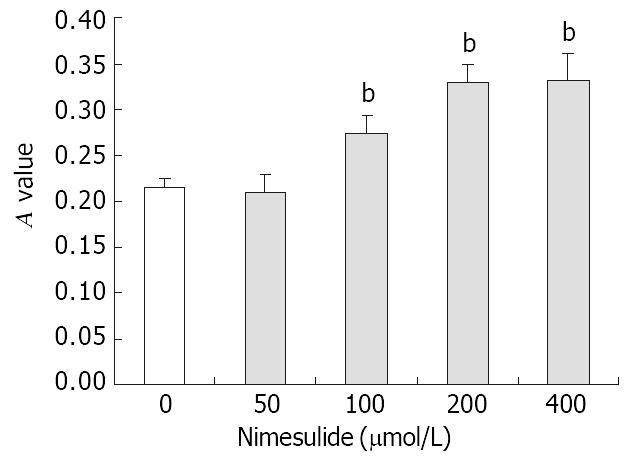
Figure 6 Effects of nimesulide on P27kip1 proteins expression in SGC-7901 cells.
The data present -x±s, n = 3. bP < 0.01 vs control.
-
Citation: Li JY, Wang XZ, Chen FL, Yu JP, Luo HS. Nimesulide inhibits proliferation
via induction of apoptosis and cell cycle arrest in human gastric adenocarcinoma cell line. World J Gastroenterol 2003; 9(5): 915-920 - URL: https://www.wjgnet.com/1007-9327/full/v9/i5/915.htm
- DOI: https://dx.doi.org/10.3748/wjg.v9.i5.915


