Copyright
©The Author(s) 2002.
World J Gastroenterol. Feb 15, 2002; 8(1): 5-12
Published online Feb 15, 2002. doi: 10.3748/wjg.v8.i1.5
Published online Feb 15, 2002. doi: 10.3748/wjg.v8.i1.5
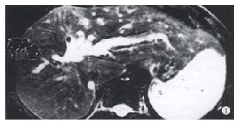
Figure 1 Hepatobiliary damages 6 years after HAE with sod.
morrhuate:MRI showed atrophy of right liver, hypertrophy of the left lateral segment and obstruc-tion of hepatic duct at the hila hepatis.
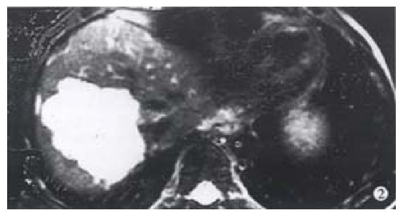
Figure 2 Hemangioma of right liver years after HAE.
The same patient. MRI showed hemangioma still in place.
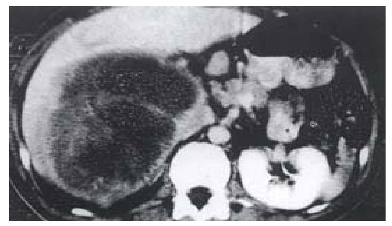
Figure 3 Neurilemmoma of the right liver: Resection of the tumr with right-sided tri-segmentectomy resulted into injury ofthe left hepatic duct.
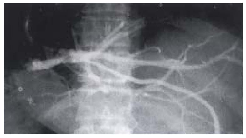
Figure 4 The same case as Fig 3.
Retrograde cholangiography of the intraphepatic duct through a PTBD catheter showing obstruction of left intrahepatic ducts and hypetrophy of the left hepatric segment.
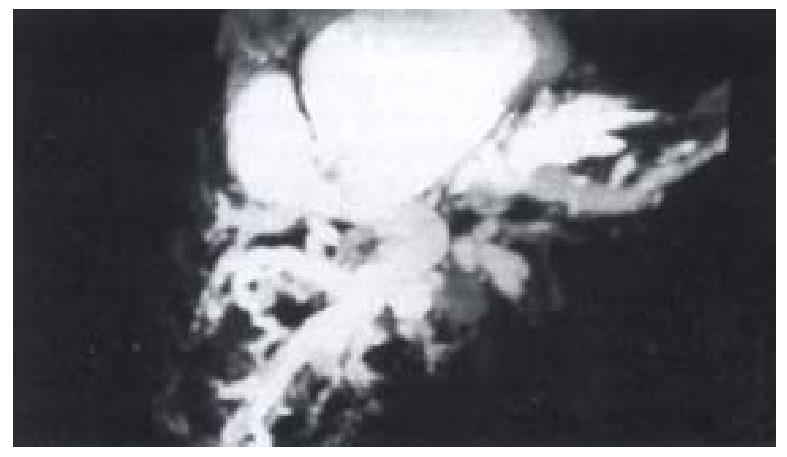
Figure 5 Bile duct injury in resection of quadrate lobe hemangioma: MRCP showing marked dilatation of the intrahepatic duct system with suphrenic collection of fluid.
Extrahepatic bile duct not visualized.
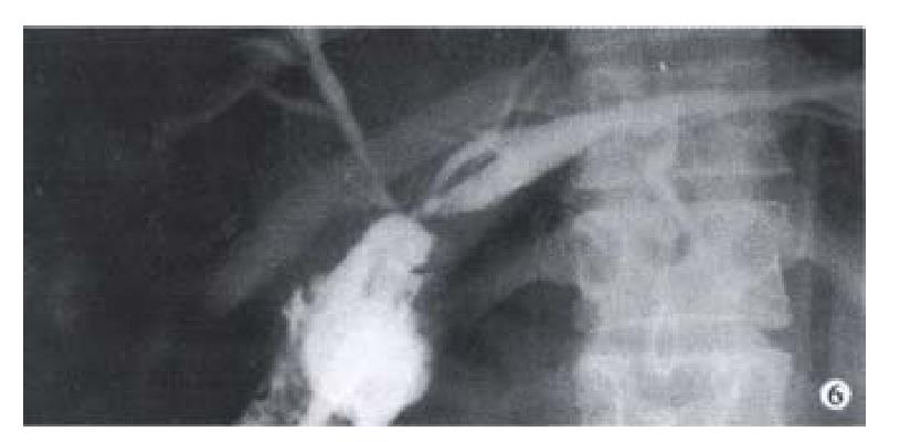
Figure 6 LC bile duct injury: Bile duct repair by hepatico-jejunostomy of Roux-en-Y tye.
Postoperative retrograde cholangiography only the right anterior segmental duct and the left hepatic duct visualized.
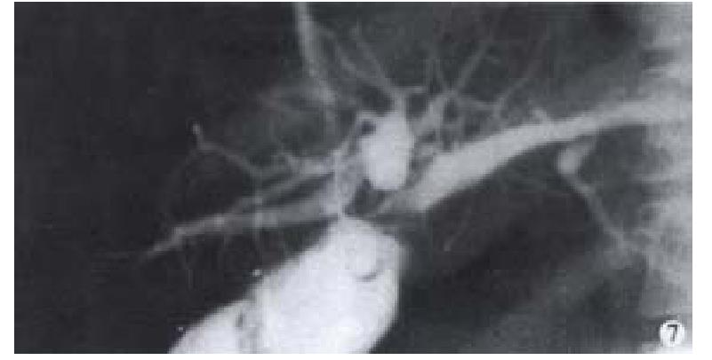
Figure 7 The same case.
PTC showing the dilated blind end of the right posterior segmental duct which was “missed” during the operation.
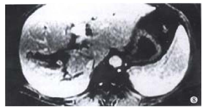
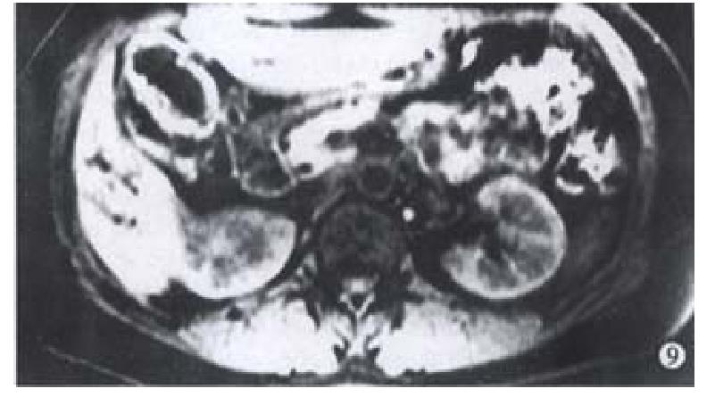
Figure 8 Fibrosis and obstruction of hepatic duct following HAE with injection of sodium morrhuate: 2 years after the procedure.
Figure 9 The same case.
Necrosis of the gallbladder and the hepatic duct.
- Citation: Huang ZQ, Huang XQ. Changing patterns of traumatic bile duct injuries: a review of forty years experience. World J Gastroenterol 2002; 8(1): 5-12
- URL: https://www.wjgnet.com/1007-9327/full/v8/i1/5.htm
- DOI: https://dx.doi.org/10.3748/wjg.v8.i1.5


