Copyright
©The Author(s) 2000.
World J Gastroenterol. Dec 15, 2000; 6(6): 812-818
Published online Dec 15, 2000. doi: 10.3748/wjg.v6.i6.812
Published online Dec 15, 2000. doi: 10.3748/wjg.v6.i6.812
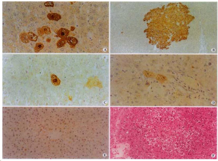
Figure 1 Immunohistochemical staining for GST-P in rat hepatic preneoplastic lesions induced by Solt-Farber protocol (A and B), in rat liver treated with DEN and AAF plus BR for 6 wk (C and D), and in normal rat liver (E).
A, GST-P-strongly positive minifoci in group B × 400; B, GST-P-strongly positive large foci in group B, × 100; C, GST-P-positive single cell in group; C, × 400; D, GST-P-weakly positive two cells in group; D, × 400; E, GST-P was negative in group A, normal rat liver, × 200; F, Hematoxilin and eosin staining for preneoplastic hepatic foci in group B, × 200.
- Citation: Yin ZZ, Jin HL, Yin XZ, Li TZ, Quan JS, Jin ZN. Effect of Boschniakia rossica on expression of GST-P, p53 and p21ras proteins in early stage of chemical hepatocarcinogenesis and its anti-inflammatory activities in rats. World J Gastroenterol 2000; 6(6): 812-818
- URL: https://www.wjgnet.com/1007-9327/full/v6/i6/812.htm
- DOI: https://dx.doi.org/10.3748/wjg.v6.i6.812


