Copyright
©The Author(s) 2024.
World J Gastroenterol. May 7, 2024; 30(17): 2311-2320
Published online May 7, 2024. doi: 10.3748/wjg.v30.i17.2311
Published online May 7, 2024. doi: 10.3748/wjg.v30.i17.2311
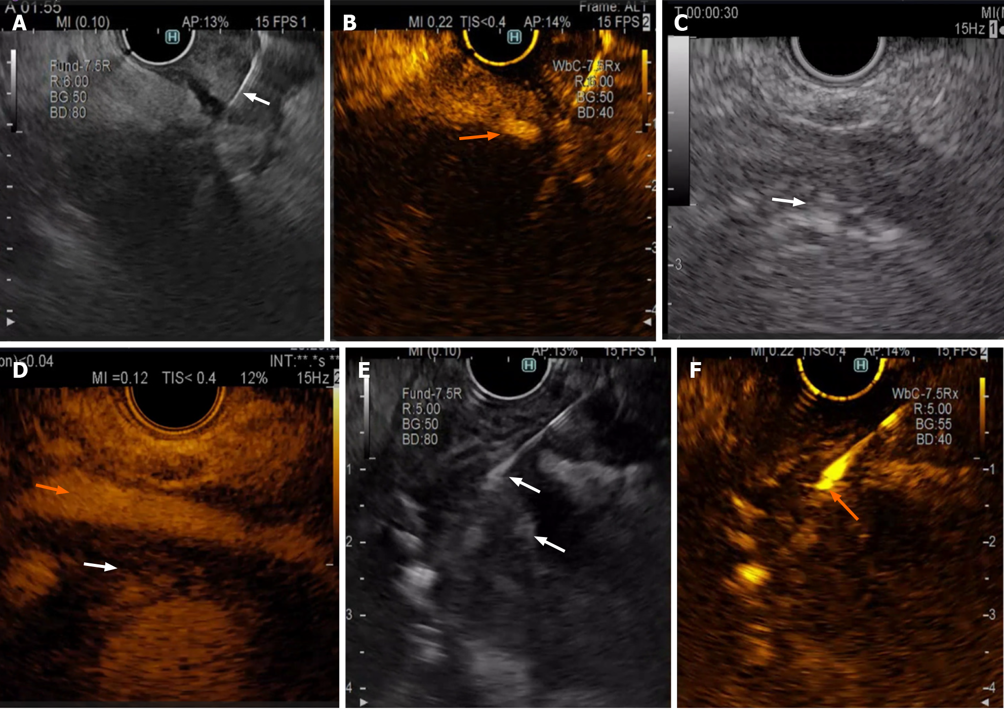
Figure 1 Contrast-enhanced endoscopic ultrasound.
A and B: Contrast-enhanced endoscopic ultrasound (CE-EUS)-guided fine needle aspiration (FNA). CE-EUS-guided FNA (white arrow) of a pancreatic mass suggestive of adenocarcinoma. A large vessel near the gut wall is more visible during contrast enhancement (orange arrow); C and D: CE-EUS view of perivascular tumour infiltration. CE-EUS view of perivascular tumour infiltration (superior mesenteric artery, orange arrow) as a hypoenhanced rim (white arrow, both images) around the artery in a patient with pancreatic head adenocarcinoma; E and F: Contrast enhanced endoscopic ultrasound-guided FNA from a hyperenhanced mural nodule inside a branch-duct intraductal papillary mucinous neoplasm (IPMN). CHEUSFNA was performed on a hyperenhanced mural nodule inside a branchduct IPMN, revealing carcinoma cells. On the left, a conventional endoscopic ultrasound image of an IPMN with multiple hyperechoic nodules in the wall (white arrows) is shown. On the right, only one of the nodules was hyperenhanced by the contrast agent, so endoscopic ultrasound-guided FNA sampling was performed.
- Citation: Gheorghiu MI, Seicean A, Pojoga C, Hagiu C, Seicean R, Sparchez Z. Contrast-enhanced guided endoscopic ultrasound procedures. World J Gastroenterol 2024; 30(17): 2311-2320
- URL: https://www.wjgnet.com/1007-9327/full/v30/i17/2311.htm
- DOI: https://dx.doi.org/10.3748/wjg.v30.i17.2311


