Copyright
©The Author(s) 1997.
World J Gastroenterol. Dec 15, 1997; 3(4): 210-212
Published online Dec 15, 1997. doi: 10.3748/wjg.v3.i4.210
Published online Dec 15, 1997. doi: 10.3748/wjg.v3.i4.210
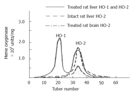
Figure 1 DEAE-Sephacel chromatography of rat liver and brain solubilized microsomes.
Solubilized microsomal fractions were loaded onto a DEAE-Sephacel column (2.5 cm × 13.6 cm) equilibrated with 20.0 mmol/L Tris-HCl buffer (pH 7.5) containing 0.05 mmol/L EDTA, 0.5% Triton X-100, 0.1% sodium cholate, and 0.05 mmol/L DTT. The column was eluted with concurrent linear gradients of KCl (0-0.4 mol/L) and Triton X-100 (0.5%-0.9%) in the same buffer. The fractions (3.0-4.0 mL) were collected and analyzed for enzyme activities.
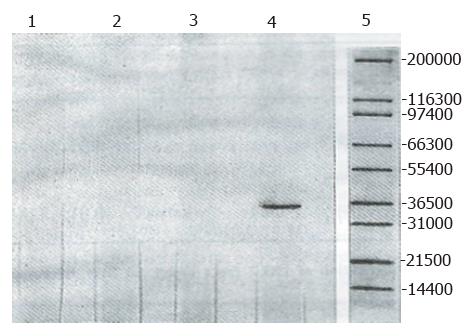
Figure 2 Western immunoblot of rat liver HO-1 and brain HO-2 preparations.
Preparations of HO-1 and HO-2 were subjected to SDS-polyacrylamide slab gel electrophoresis, electroblotted onto nitrocellulose membranes and visualized as described in the “experimental procedures”. 1 and 3, controls; 2, liver HO-1; 4, brain HO-2; 5, molecular weight markers.
- Citation: Xia ZW, Li YZ, Chen SN, Shen QX, Ben XM, Yu SC. Analysis of two constitutive forms of microsomal heme oxygenase in different rat tissues. World J Gastroenterol 1997; 3(4): 210-212
- URL: https://www.wjgnet.com/1007-9327/full/v3/i4/210.htm
- DOI: https://dx.doi.org/10.3748/wjg.v3.i4.210


