Copyright
©The Author(s) 2021.
World J Gastroenterol. Oct 7, 2021; 27(37): 6322-6331
Published online Oct 7, 2021. doi: 10.3748/wjg.v27.i37.6322
Published online Oct 7, 2021. doi: 10.3748/wjg.v27.i37.6322
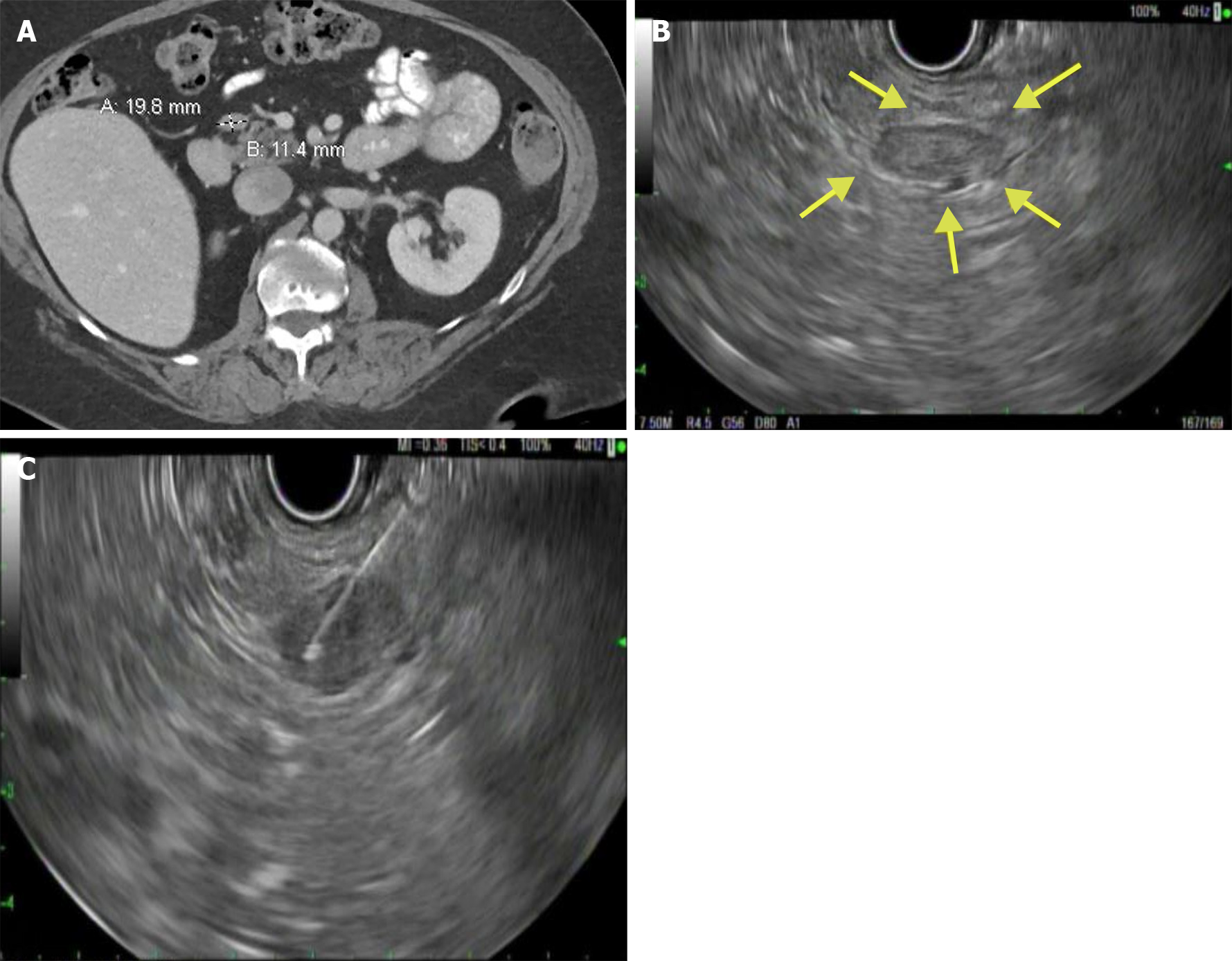
Figure 1 Computed tomography and endoscopy examinations.
A: Computed tomography of abdomen pelvis showing a peripancreatic lymph node adjacent to the pancreatic head; B: Endoscopic ultrasonography showing a hypoechoic lesion near the pancreatic head; C: Endoscopic ultrasound-guided fine needle aspiration of the peripancreatic lesion.
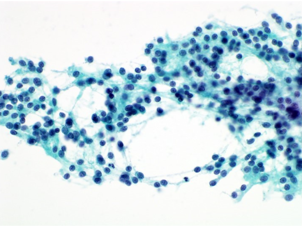
Figure 2 Fine needle aspiration direct smear.
Papanicolaou stain, × 400.
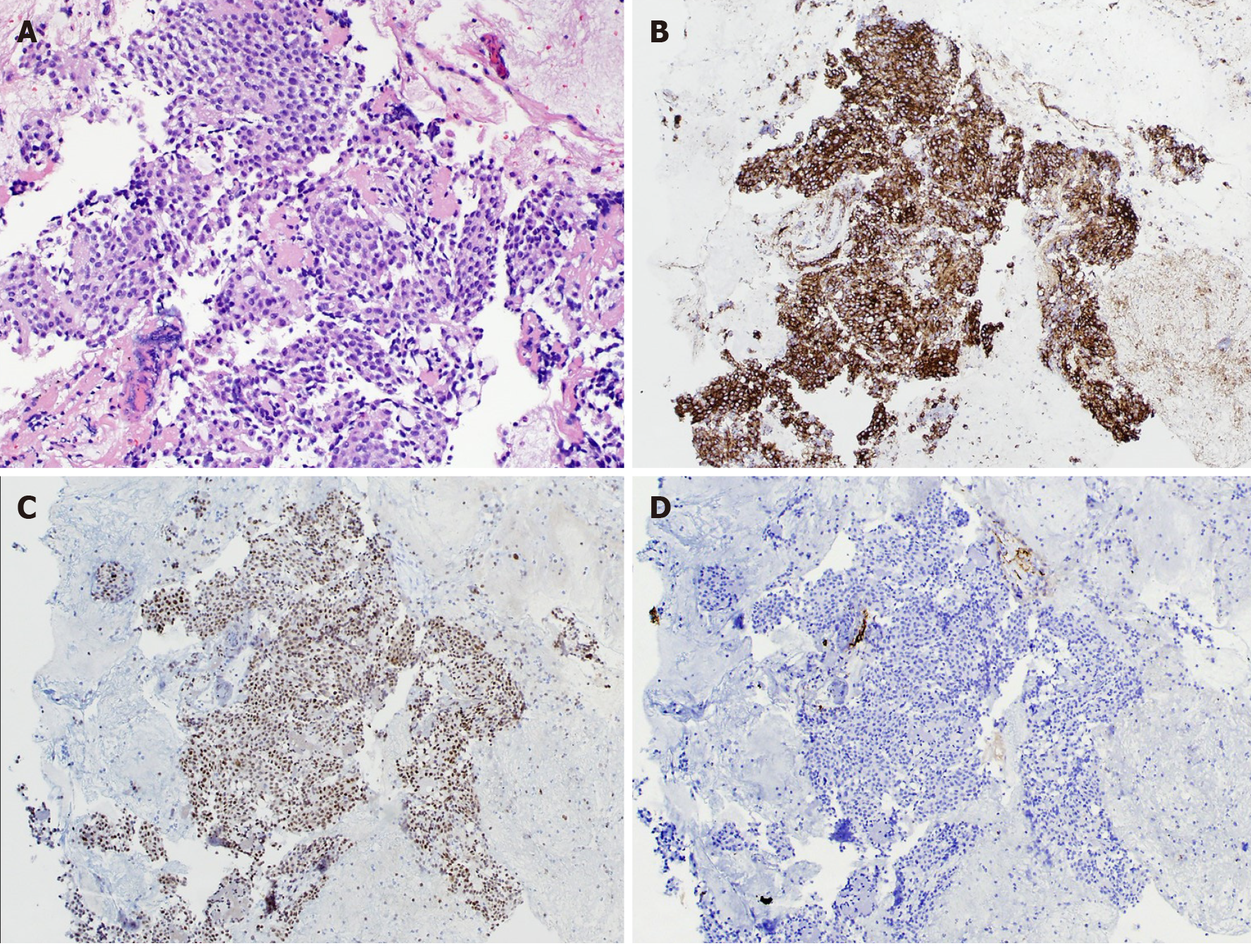
Figure 3 Fine needle aspiration cell block.
A: Hematoxylin and eosin stain, × 200; B: Diffuse cytoplasmic staining, chromogranin (× 100); C: Diffuse nuclear staining, GATA-3, (× 100); D: No staining, keratin cocktail (× 100).
- Citation: Lanke G, Stewart JM, Lee JH. Pancreatic paraganglioma diagnosed by endoscopic ultrasound-guided fine needle aspiration: A case report and review of literature. World J Gastroenterol 2021; 27(37): 6322-6331
- URL: https://www.wjgnet.com/1007-9327/full/v27/i37/6322.htm
- DOI: https://dx.doi.org/10.3748/wjg.v27.i37.6322


