Copyright
©The Author(s) 2021.
World J Gastroenterol. Aug 7, 2021; 27(29): 4929-4938
Published online Aug 7, 2021. doi: 10.3748/wjg.v27.i29.4929
Published online Aug 7, 2021. doi: 10.3748/wjg.v27.i29.4929
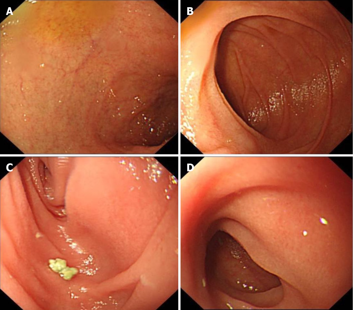
Figure 1 Endoscopic images at the onset of postcolectomy enteritis in 2016.
A: Gastroscopy showing a normal duodenal bulb; B: Gastroscopy showing a normal descending duodenum; C and D: Endoscopy with loop ileostomy showing a normal appearance of the neoileum.
Figure 2 Pathological findings at the onset of postcolectomy enteritis in 2016.
A: The biopsy from the duodenum showed chronic inflammation, moderate villous atrophy, and decreased goblet cells (× 40); B: Active inflammation, cryptitis, and destroyed crypts were observed in some areas (× 100).
Figure 3 Endoscopic images at the onset of autoimmune enteropathy in 2019.
A: Gastroscopy showing diffuse villous atrophy in the mucosa of the duodenal bulb; B and C: Gastroscopy showing diffuse villous atrophy in the mucosa of the descending duodenum; D and E: Enteroscopy showing diffuse villous atrophy in the mucosa of the ileum; F and G: Enteroscopy showing diffuse villous atrophy in the mucosa of the ileal pouch.
Figure 4 Pathological findings at the onset of autoimmune enteropathy in 2019.
The biopsy showed prominent chronic inflammation, flat villi, and a lack of goblet and Paneth cells (A), and apoptosis bodies were found (arrow) (B).
- Citation: Zhou QY, Zhou WX, Sun XY, Wu B, Zheng WY, Li Y, Qian JM. Autoimmune enteropathy and primary biliary cholangitis after proctocolectomy for ulcerative colitis: A case report and review of the literature. World J Gastroenterol 2021; 27(29): 4929-4938
- URL: https://www.wjgnet.com/1007-9327/full/v27/i29/4929.htm
- DOI: https://dx.doi.org/10.3748/wjg.v27.i29.4929


