Copyright
©The Author(s) 2018.
World J Gastroenterol. Oct 7, 2018; 24(37): 4291-4296
Published online Oct 7, 2018. doi: 10.3748/wjg.v24.i37.4291
Published online Oct 7, 2018. doi: 10.3748/wjg.v24.i37.4291
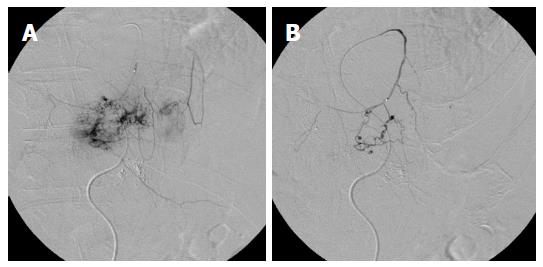
Figure 1 Angiography.
A: Selective angiography of the left inferior phrenic artery shows feeding of the anterior part of the tumor without a shunt to the pulmonary vasculature; B: Angiography during glue embolization shows no influx into the pulmonary vasculature.
Figure 2 Infused 140 mg of miriplatin suspended in 7 mL of lipiodol via the left hepatic artery.
A: Left hepatic angiography shows a large hypervascular tumor in the left hepatic lobe and intrahepatic metastases neighborhood without an intratumoral AV shunt; B: Repeated left hepatic angiography shows a vascular lake in the superior portion of the tumor (arrow) that developed after chemoembolization; C: Venous phase shows drainage into the pericardiacophrenic vein (arrow heads), which was unrecognized until remasking and pixel shifting were performed. AV: Arteriovenous.
Figure 3 Non-contrast brain computed tomography scan obtained 30 min after chemoembolization shows multiple increased attenuated lesions in the bilateral cerebral hemisphere, consistent with lipiodol embolism.
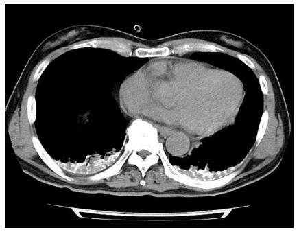
Figure 4 Chest computed tomography scan obtained 30 min after chemoembolization shows lipiodol deposition at both lung bases.
- Citation: Ishimaru H, Morikawa M, Sakugawa T, Sakamoto I, Motoyoshi Y, Ikebe Y, Uetani M. Cerebral lipiodol embolism related to a vascular lake during chemoembolization in hepatocellular carcinoma: A case report and review of the literature. World J Gastroenterol 2018; 24(37): 4291-4296
- URL: https://www.wjgnet.com/1007-9327/full/v24/i37/4291.htm
- DOI: https://dx.doi.org/10.3748/wjg.v24.i37.4291


