Copyright
©The Author(s) 2017.
World J Gastroenterol. Feb 7, 2017; 23(5): 859-868
Published online Feb 7, 2017. doi: 10.3748/wjg.v23.i5.859
Published online Feb 7, 2017. doi: 10.3748/wjg.v23.i5.859
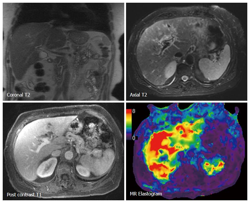
Figure 1 Magnetic resonance elastography in untreated autoimmune hepatitis.
An 84-year-old female with grade 4 inflammation and cirrhosis. The liver has normal contour with no morphological features of cirrhosis. Lab tests were: AST 473, ALT 406, APRI 6.26 and FIB-4 10.31. LS was 6.4 kPa consistent with cirrhosis. ALT: Alanine aminotransferase; AST: Aspartate aminotransferase; APRI: AST to platelet ratio index; FIB-4: Fibrosis 4 test; LS: Liver stiffness.
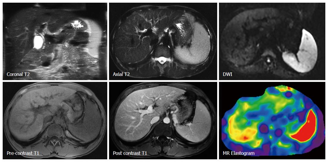
Figure 2 Magnetic resonance elastography in treated autoimmune hepatitis.
A 43-year-old male with grade 2 inflammation and advanced fibrosis. MRI images show no features to suggest advanced fibrosis. Note prominent spleen. Lab tests were AST 81, ALT 147, FIB-4 2.95 and APRI 1.98. LS was 5.1 kPa consistent with advanced fibrosis. ALT: Alanine aminotransferase; AST: Aspartate aminotransferase; APRI: AST to platelet ratio index; FIB-4: Fibrosis 4 test; LS: Liver stiffness.
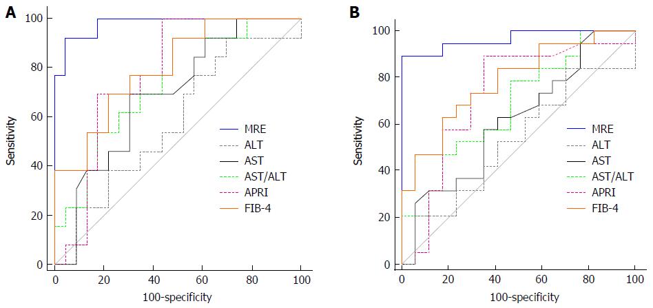
Figure 3 Graph showing area under the receiver operating characteristic curves of magnetic resonance elastography and lab tests for prediction of advanced fibrosis (A) and cirrhosis (B) in autoimmune hepatitis.
- Citation: Wang J, Malik N, Yin M, Smyrk TC, Czaja AJ, Ehman RL, Venkatesh SK. Magnetic resonance elastography is accurate in detecting advanced fibrosis in autoimmune hepatitis. World J Gastroenterol 2017; 23(5): 859-868
- URL: https://www.wjgnet.com/1007-9327/full/v23/i5/859.htm
- DOI: https://dx.doi.org/10.3748/wjg.v23.i5.859


