Copyright
©The Author(s) 2017.
World J Gastroenterol. Nov 21, 2017; 23(43): 7807-7812
Published online Nov 21, 2017. doi: 10.3748/wjg.v23.i43.7807
Published online Nov 21, 2017. doi: 10.3748/wjg.v23.i43.7807
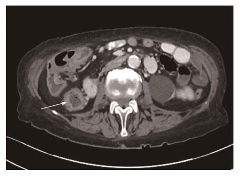
Figure 1 Axial image from computed tomography Chest/Abdomen/Pelvis performed with intravenous (IV) and oral contrast showing non-specific colonic wall thickening in the ascending colon (arrowed) and hepatic flexure characterised by discontinuous mucosal hyperenhancement and submucosal oedema reflecting ulceration and inflammation.
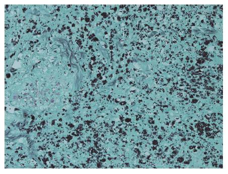
Figure 2 Extensive infiltration of bowel mucosa by the small histoplasmosis, Grocott stain.
- Citation: Agrawal N, Jones DE, Dyson JK, Hoare T, Melmore SA, Needham S, Thompson NP. Fatal gastrointestinal histoplasmosis 15 years after orthotopic liver transplantation. World J Gastroenterol 2017; 23(43): 7807-7812
- URL: https://www.wjgnet.com/1007-9327/full/v23/i43/7807.htm
- DOI: https://dx.doi.org/10.3748/wjg.v23.i43.7807


