Copyright
©The Author(s) 2017.
World J Gastroenterol. Jun 21, 2017; 23(23): 4252-4261
Published online Jun 21, 2017. doi: 10.3748/wjg.v23.i23.4252
Published online Jun 21, 2017. doi: 10.3748/wjg.v23.i23.4252
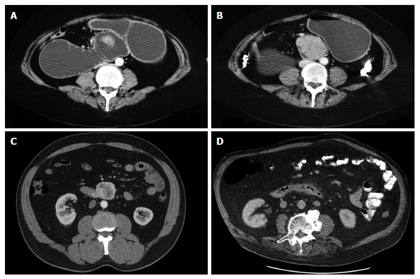
Figure 1 Representative axial computed tomography imaging of duodenal adenocarcinoma.
A and B: obstruction due to a large duodenal mass (same patient); C: exophytic mass without obstruction; D: subtle thickening of duodenum and periduodenal fat stranding reported as duodenitis, but in fact was a malignant tumour on post resection histology.
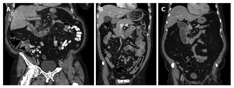
Figure 2 Representative coronal computed tomography imaging of duodenal adenocarcinoma.
A: thickening of the duodenal wall with non-obstructive narrowing; B: mass in the distal duodenum, C: stricture of D2/3 junction with some obstructive features.
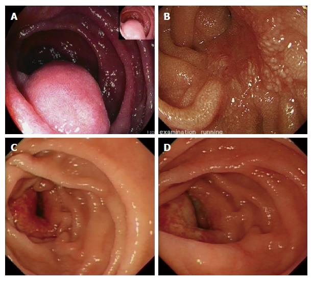
Figure 3 Endoscopic features.
A: pedunculated lesion (gastrointestinal stromal tumour); B: sessile lesion (large adenoma with previous endoscopic mucosal resection); C and D: malignant ulceration of a duodenal adenocarcinoma.
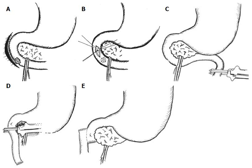
Figure 4 Operative technique.
After wide Kocherisation (A) the papilla and lesion are palpated. For benign pathology close to the papilla, the ampullary complex can be further protected by cannulation (B). The proximal jejunum is transected (C), the distal duodenum taken off its short vessels and the resection is completed (D). Reconstruction is by a retrocolic isoperistaltic side-to-side duodenojejunostomy (E).
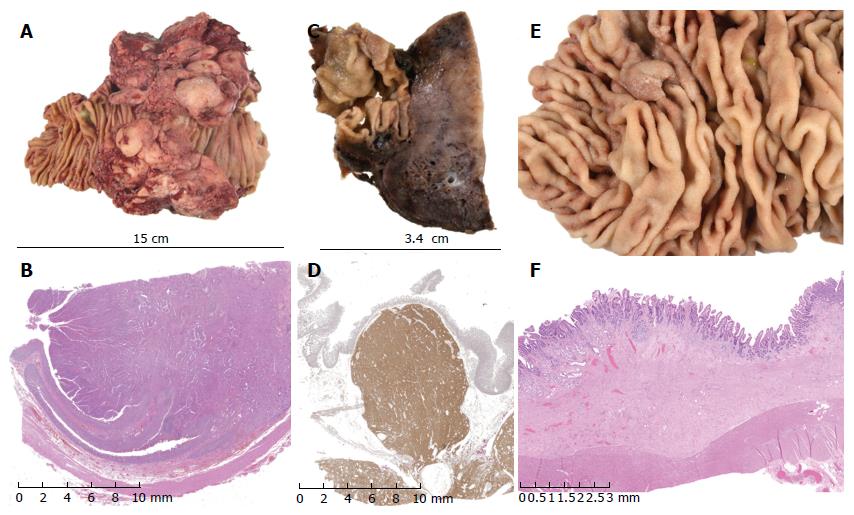
Figure 5 Pathological findings.
A and B: exophytic lesion in the duodenum shown to be a moderately differentiated duodenal adenocarcinoma on histology (haematoxylin and eosin stain); C and D: gastrointestinal stromal tumour confirmed on immunohistochemistry with CD117 and DOG1 staining; E and F: tubulovillous adenoma of the duodenum with low-grade dysplasia on histology (haematoxylin and eosin stain).
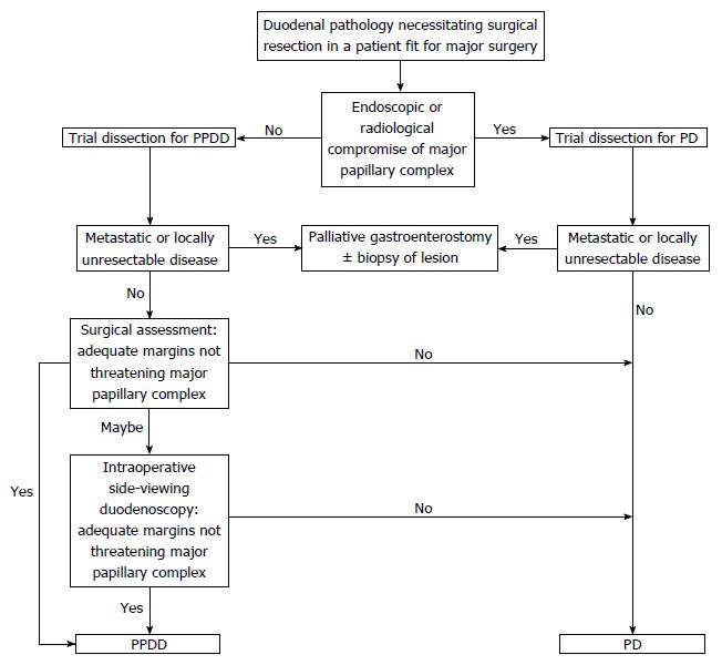
Figure 6 Flow chart summarising the local algorithm for the management of infrapapillary duodenal lesions.
PD: Pancreaticoduodenectomy; PPDD: Pancreas preserving distal duodenectomy.
- Citation: Mitchell WK, Thomas PF, Zaitoun AM, Brooks AJ, Lobo DN. Pancreas preserving distal duodenectomy: A versatile operation for a range of infra-papillary pathologies. World J Gastroenterol 2017; 23(23): 4252-4261
- URL: https://www.wjgnet.com/1007-9327/full/v23/i23/4252.htm
- DOI: https://dx.doi.org/10.3748/wjg.v23.i23.4252


