Copyright
©The Author(s) 2017.
World J Gastroenterol. Mar 14, 2017; 23(10): 1828-1835
Published online Mar 14, 2017. doi: 10.3748/wjg.v23.i10.1828
Published online Mar 14, 2017. doi: 10.3748/wjg.v23.i10.1828
Figure 1 Porcine model creation process.
A: Intramuscular injection with anesthetic Zoteil 50 at 1 mL/kg; B: A 0.5 cm minimal incision made in the anus at 3, 9, and 12 o’clock position and ligated rubber used; C: Directly cross through the anal sphincter with rubber band seton.
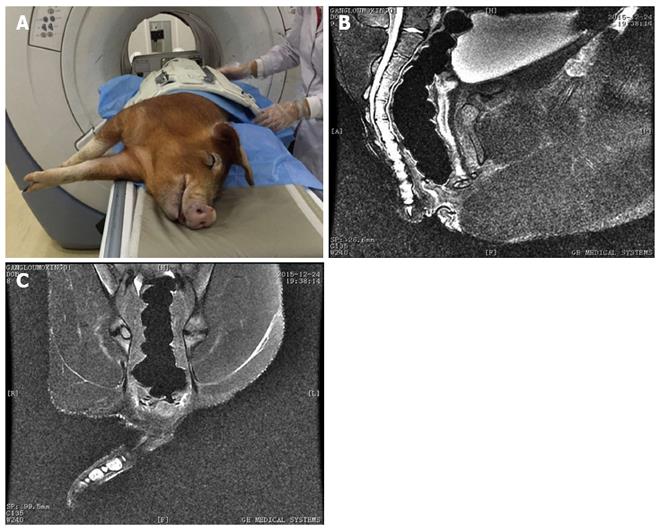
Figure 2 Magnetic resonance imaging evaluation of created fistulas.
A: Created fistulas; B and C: Typical fistula imaging was found and it was very similar to human anal fistula imaging of magnetic resonance imaging.
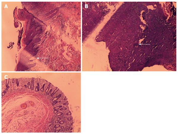
Figure 3 Pathological findings of created fistulas.
A: Epithelization in the created fistula tract; B and C: Epithelization and granulation were found in the created fistula tract.
- Citation: A Ba-Bai-Ke-Re MMTJ, Chen H, Liu X, Wang YH. Experimental porcine model of complex fistula-in-ano. World J Gastroenterol 2017; 23(10): 1828-1835
- URL: https://www.wjgnet.com/1007-9327/full/v23/i10/1828.htm
- DOI: https://dx.doi.org/10.3748/wjg.v23.i10.1828


