Copyright
©The Author(s) 2017.
World J Gastroenterol. Jan 7, 2017; 23(1): 110-120
Published online Jan 7, 2017. doi: 10.3748/wjg.v23.i1.110
Published online Jan 7, 2017. doi: 10.3748/wjg.v23.i1.110
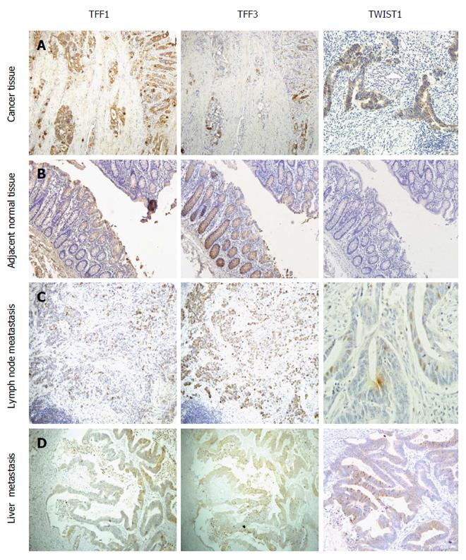
Figure 1 TFF1, TFF3 and TWIST1 expression trends in colorectal cancer (A), adjacent normal tissue (B), metastatic lymph node (C), and liver metastasis (D).
Compared with normal mucosal tissue, colorectal cancer tissues expressed significantly higher TFF1 and TWIST1 and lower TFF3. Compared to primary colorectal cancer tissue, there were no statistical differences in TFF3 and TWIST1 expression in metastatic lymph node or liver metastatic tissue. But disparity in TFF1 expression between metastatic lymph node and primary colorectal cancer had statistical significance.
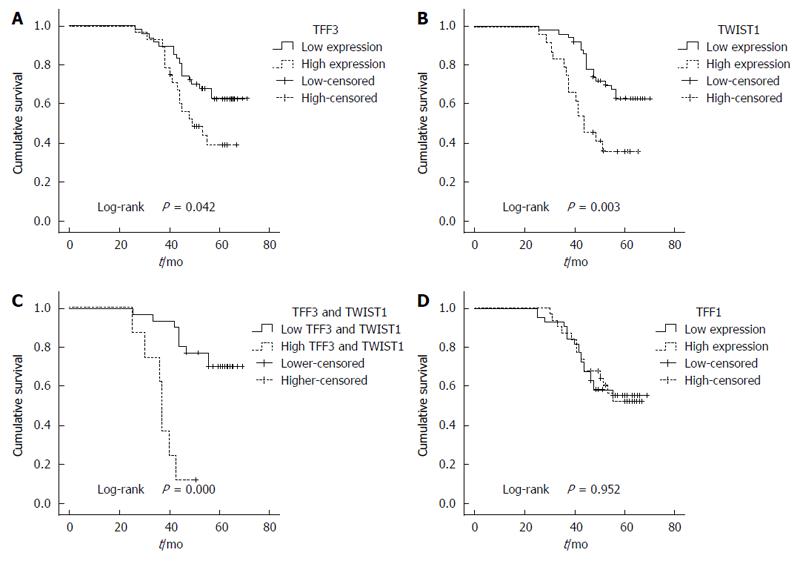
Figure 2 Correlation between protein expression in colorectal cancer and overall survival.
The survival analysis indicated that the overall survival of the patients with higher TFF3 or TWIST1 expression was significantly less than that of the patients with lower TFF3 or TWIST1 expression (A and B); Higher expression of the TFF3 and TWIST1 correlated with even worse overall survival (C); TFF1 expression had no correlation with the survival (D).
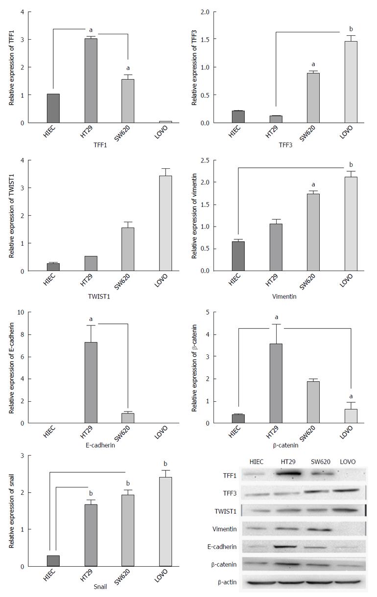
Figure 3 The mRNA and protein expression of TFF1, TFF3, TWIST1, E-cadherin, vimentin, and β-catenin in colon cancer cell lines with different invasion potentials.
The expression of TFF3 and TWIST1 in cancer cell lines was higher than that in normal human intestinal epithelial cell line, and the expression intensity was inclined to rise with the increase in metastatic potential both at protein and mRNA levels. But TFF1 expression had the opposite tendency. The TFF1 protein and mRNA expression decreased and TWIST1 and TFF3 expression increased gradually with the increase in metastatic potential of cell lines. E-cadherin and β-catenin expression tended to decrease, while vimentin and TWIST1 expression was inclined to rise with the increase in metastatic potential. aP < 0.05, bP < 0.01.
- Citation: Yusup A, Huji B, Fang C, Wang F, Dadihan T, Wang HJ, Upur H. Expression of trefoil factors and TWIST1 in colorectal cancer and their correlation with metastatic potential and prognosis. World J Gastroenterol 2017; 23(1): 110-120
- URL: https://www.wjgnet.com/1007-9327/full/v23/i1/110.htm
- DOI: https://dx.doi.org/10.3748/wjg.v23.i1.110


