Copyright
©The Author(s) 2016.
World J Gastroenterol. Jun 7, 2016; 22(21): 5132-5136
Published online Jun 7, 2016. doi: 10.3748/wjg.v22.i21.5132
Published online Jun 7, 2016. doi: 10.3748/wjg.v22.i21.5132
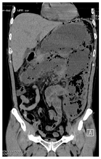
Figure 1 Abdominal computed tomography scan showing a huge multilocular walled-off necrosis replacing the body and tail of the pancreas, which extended to the pelvis.
Gas bubbles were observed in the cavity.
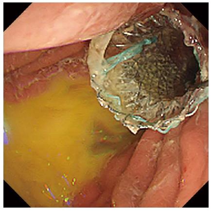
Figure 2 Successful deployment of a wide-caliber fully covered TTS Niti-S esophageal stent.
Purulent fluid was observed in the gastric lumen.
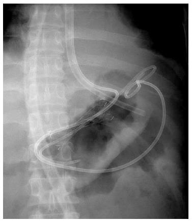
Figure 3 A 7-Fr double-pigtail plastic stent and a 7-Fr nasocystic catheter were deployed through the fully covered metal stent.
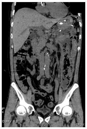
Figure 4 Computed tomography one week after initial drainage showed an undrained subcavity, located mainly at the left anterior pararenal space that extended to the left pelvis.
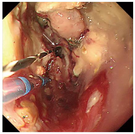
Figure 5 Endoscopic view of the cavity of walled-off necrosis by a modified single transluminal gateway transcystic multiple drainage technique.
An upper endoscope was inserted into the walled-off necrosis (WON) through the fistula and a narrow connection route within the main cavity to the subcavity could be identified directly (white arrow).
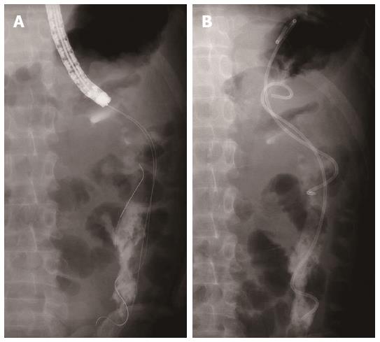
Figure 6 Fluoroscopic view of modified single transluminal gateway transcystic multiple drainage technique.
With a direct view of the connection route, a 0.025-inch guidewire was inserted into the subcavity (A) and two 7-Fr double-pigtail plastic stents were deployed (B).
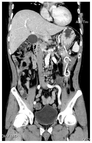
Figure 7 Follow-up computed tomography obtained one month after discharge revealed the WON had mostly collapsed.
- Citation: Minaga K, Kitano M, Imai H, Yamao K, Kamata K, Miyata T, Matsuda T, Omoto S, Kadosaka K, Yoshikawa T, Kudo M. Modified single transluminal gateway transcystic multiple drainage technique for a huge infected walled-off pancreatic necrosis: A case report. World J Gastroenterol 2016; 22(21): 5132-5136
- URL: https://www.wjgnet.com/1007-9327/full/v22/i21/5132.htm
- DOI: https://dx.doi.org/10.3748/wjg.v22.i21.5132


