Copyright
©The Author(s) 2016.
World J Gastroenterol. Mar 14, 2016; 22(10): 2993-3005
Published online Mar 14, 2016. doi: 10.3748/wjg.v22.i10.2993
Published online Mar 14, 2016. doi: 10.3748/wjg.v22.i10.2993
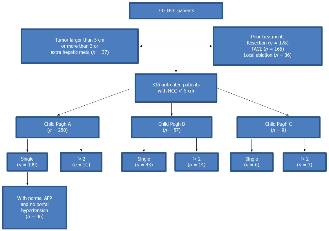
Figure 1 Flow chart summarizing the patients selected for this study.
HCC: Hepatocellular carcinoma; TACE: Transcatheter arterial chemoembolization; AFP: Alpha-fetoprotein.
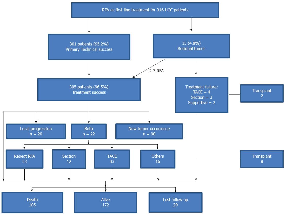
Figure 2 Outcomes of radiofrequency ablation as a first-line treatment for 316 patients with liver cirrhosis and hepatocellular carcinoma.
RFA: Radiofrequency ablation; HCC: Hepatocellular carcinoma; TACE: Transcatheter arterial chemoembolization.
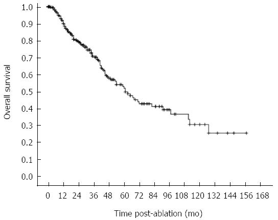
Figure 3 Kaplan-Meier curve showing an overall 10-year survival.
Overall estimated 1-, 3-, 5-, 7-, and 10-year survival rates are 90.0%, 70.8%, 49.7%, 41.1%, and 28.4%, respectively (n = 234, 181, 136, and 71 for 3, 5, 7, and 10 years, respectively).
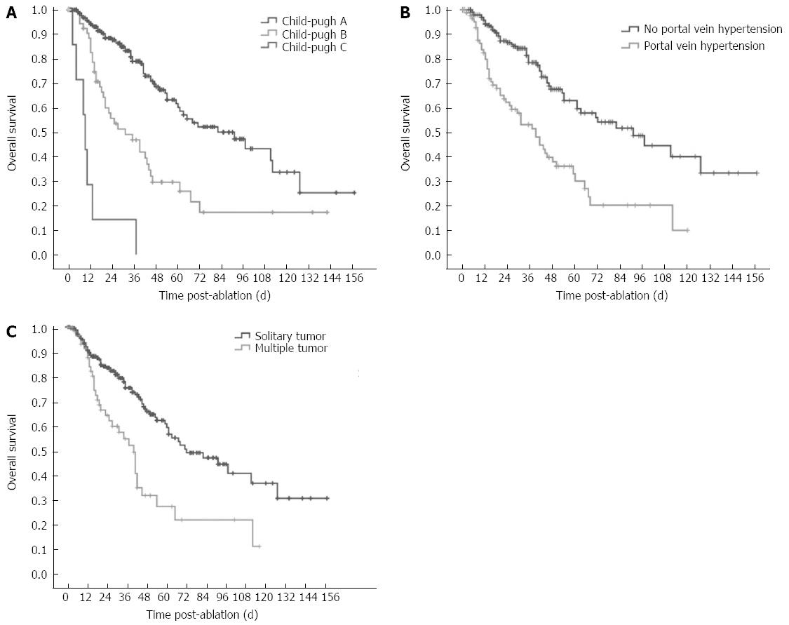
Figure 4 Post-radiofrequency ablation overall survival curves in hepatocellular carcinoma patients with different independent prognostic factors.
A: The 1-, 3-, 5-, 7-, and 10-year overall survival for patients with Child-Pugh class A liver function of were 94.1%, 78.9%, 60.1%, 50.5%, and 33.6%, whereas the overall survival rates were 82.5%, 46.8%, 25.9%, 17.2%, and 17.2%, respectively, for Child-Pugh class B, and 28.6%, 14.3%, 0%, 0%, and 0% for Child-Pugh class C, respectively (P < 0.001); B: The 1-, 3-, 5-, 7-, and 10-year overall survival rates were 94.2%, 78.5%, 63.0%, 51.8%, and 36.2%, respectively, for patients without portal vein hypertension and 79.8%, 53.2%, 30.2%, 20.4%, and 10.2%, respectively, for patients with portal vein hypertension (P = 0.002); C: The 1-, 3-, 5-, 7-, and 10-year survival rates were 90.6%, 75.3%, 59.3%, 46.8%, and 34.5%, respectively, for patients with a single tumor and 87.6%, 54.6%, 27.0%, 21.6%, and 10.8%, respectively, for patients with more than one tumor (P = 0.003).
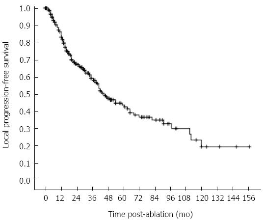
Figure 5 Kaplan-Meier curve shows a local progression-free 10-year survival.
The 1-, 3-, 5-, 7- and 10-year local progression-free survival rates were 83.2%, 59.3%, 42.7%, 35.1%, and 19.5%, respectively.
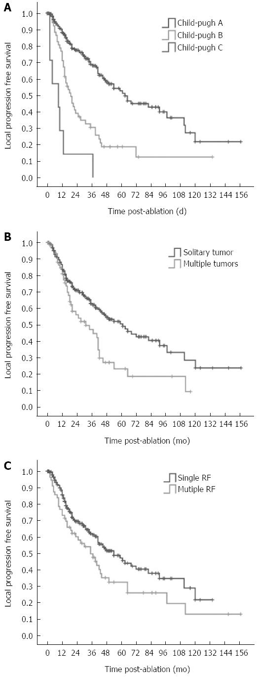
Figure 6 Post-radiofrequency ablation local progression-free survival curves in hepatocellular carcinoma patients with different independent prognostic factors.
A: Patients with Child-Pugh class A had significantly greater local progression-free survival rate than patients with Child-Pugh class B and C (P < 0.001); B: Patients with a single tumor had a significantly higher local progression-free survival rate than patients with more tumors (P = 0.036); C: Patients who had one radiofrequency ablation session had a significantly greater local progression-free survival rate than patients who had multiple sessions (P = 0.002).
- Citation: Yang W, Yan K, Goldberg SN, Ahmed M, Lee JC, Wu W, Zhang ZY, Wang S, Chen MH. Ten-year survival of hepatocellular carcinoma patients undergoing radiofrequency ablation as a first-line treatment. World J Gastroenterol 2016; 22(10): 2993-3005
- URL: https://www.wjgnet.com/1007-9327/full/v22/i10/2993.htm
- DOI: https://dx.doi.org/10.3748/wjg.v22.i10.2993


