Copyright
©2014 Baishideng Publishing Group Co.
World J Gastroenterol. Feb 7, 2014; 20(5): 1325-1331
Published online Feb 7, 2014. doi: 10.3748/wjg.v20.i5.1325
Published online Feb 7, 2014. doi: 10.3748/wjg.v20.i5.1325
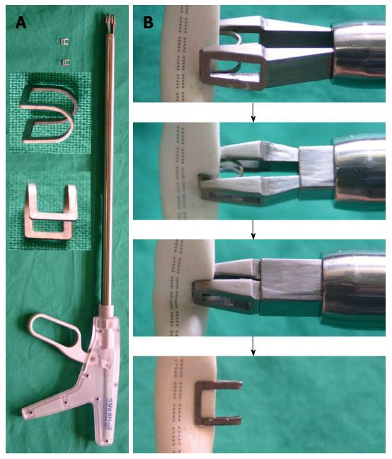
Figure 1 Proposed laparoscopic hemostatic clip and clip applicator.
A: Shape of the clip and clip applier; B: Procedure for sealing a damaged vessel without fully occluding it.
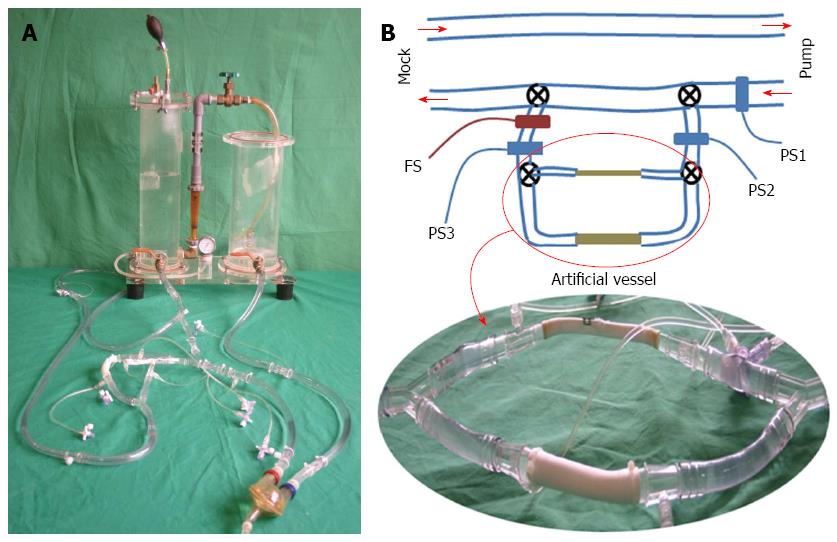
Figure 2 Setting for in vitro evaluation of the clip.
A: Closed-flow loop including a pulsatile pump, a mock circulatory device, and 2 artificial vessels; B: 1/4-inch and 1/2-inch artificial vessels. PS: Pressure sensor; FS: Flow sensor.
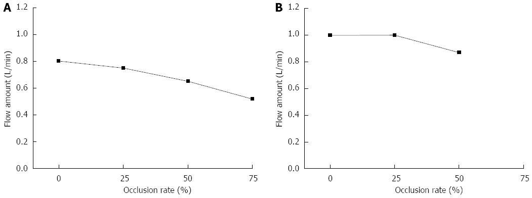
Figure 3 Flow variations according to the occlusion rate.
A: 1/4-inch artificial vessel; B: 1/2-inch artificial vessel.
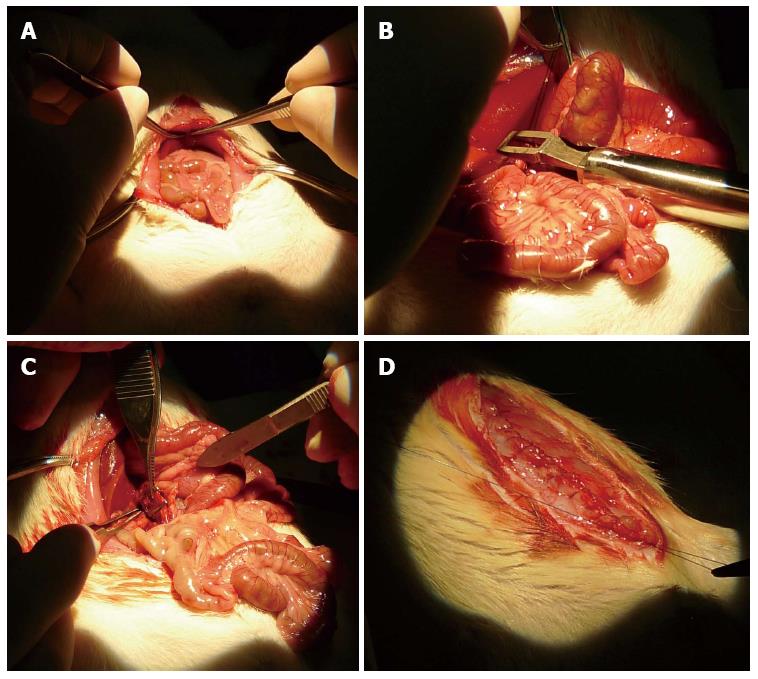
Figure 4 In vivo experiments for full-occlusion testing.
A: The rat is supine on the operating table with the abdomen opened; B: The clip applier is positioned on the target vessel; C: The target vessel is clipped; D: The abdomen is sutured after clipping.
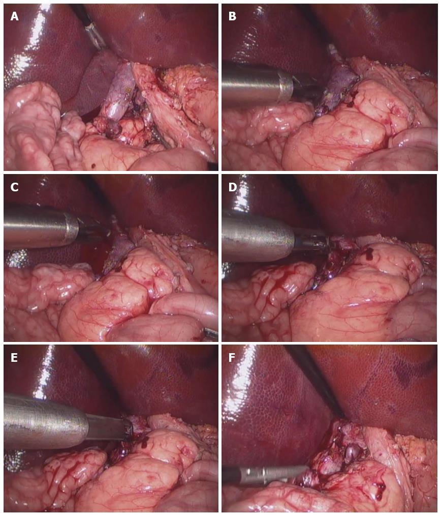
Figure 5 In vivo experiments for partial-occlusion testing.
A: Separating the target vessel (infra-hepatic inferior vena cava) from surrounding tissues; B: Scratching the surface of the target vessel using electrocautery; C: Inducing intentional bleeding on the target vessel; D: Grabbing the bleeding site using the clip applier; E: Stanching the bleeding site on the target vessel by implanting the proposed hemostatic clip; F: Suctioning the blood around the target vessel after hemostasis.
Figure 6 Measurements of blood flow through the clipped inferior vena cava using an ultrasonic Doppler machine.
The probe was applied to the positions before (A), on (B), and after the clipped site (C), respectively. The Doppler signal was slightly increased at the clipped site because the diameter of the vessel decreased after clipping the bleeding site.
- Citation: Nam KW, Lee SB, Kim IY, Kim KG, Park SJ. A new hemostatic clip for endoscopic surgery that can maintain blood flow after clipping. World J Gastroenterol 2014; 20(5): 1325-1331
- URL: https://www.wjgnet.com/1007-9327/full/v20/i5/1325.htm
- DOI: https://dx.doi.org/10.3748/wjg.v20.i5.1325


