Copyright
©2014 Baishideng Publishing Group Inc.
World J Gastroenterol. Sep 7, 2014; 20(33): 11815-11825
Published online Sep 7, 2014. doi: 10.3748/wjg.v20.i33.11815
Published online Sep 7, 2014. doi: 10.3748/wjg.v20.i33.11815
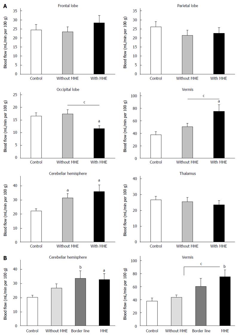
Figure 1 Blood flow in the different brain regions studied.
A: Blood flow in frontal lobe, parietal lobe, occipital lobe, vermis, cerebellar hemisphere and thalamus in controls and cirrhotic patients without and with minimal hepatic encephalopathy (MHE); B: Blood flow in cerebellar hemisphere and vermis including the patients (n = 7) showing Psychometric Hepatic Encephalopathy Score (PHES) = -3, considered as borderline. Data are expressed as mL of blood per minute per 100 g of brain tissue (mean ± SEM) of 14 controls, 24 patients without and 16 with MHE. aP < 0.05; bP < 0.01 vs control, Values significantly different between patients with and without MHE are indicated by cP < 0.05.
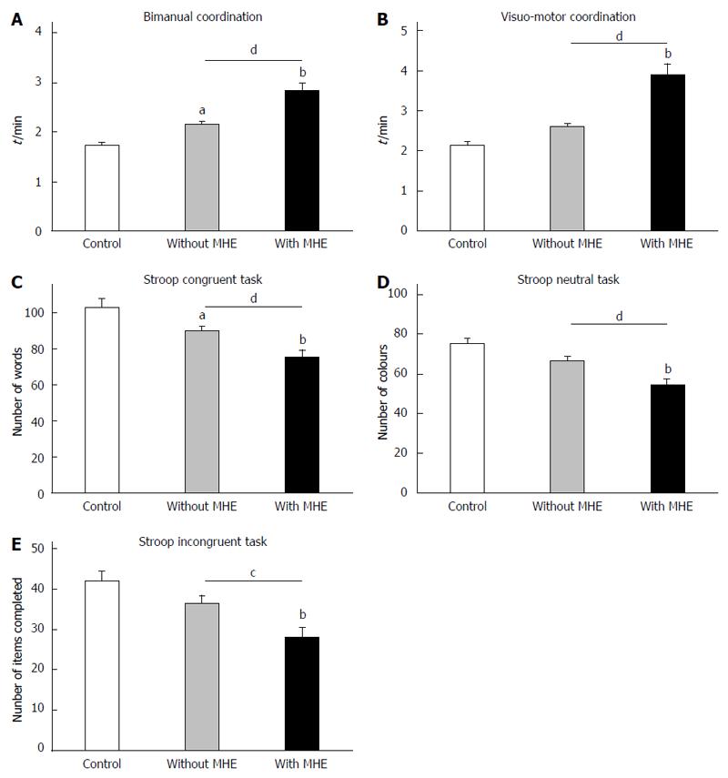
Figure 2 Performance in the Stroop, bimanual and visuo-motor coordination tests.
A: Bimanual coordination test; B: Visuo-motor coordination test; C: Stroop congruent task; D: Stroop neutral task; E: Stroop incongruent task. Values are the mean ± SEM of 14 controls, 24 patients without and 16 with MHE. aP < 0.05; bP < 0.01 vs control, Values significantly different between patients with and without MHE are indicated by cP < 0.05; dP < 0.01.
- Citation: Felipo V, Urios A, Giménez-Garzó C, Cauli O, Andrés-Costa MJ, González O, Serra MA, Sánchez-González J, Aliaga R, Giner-Durán R, Belloch V, Montoliu C. Non invasive blood flow measurement in cerebellum detects minimal hepatic encephalopathy earlier than psychometric tests. World J Gastroenterol 2014; 20(33): 11815-11825
- URL: https://www.wjgnet.com/1007-9327/full/v20/i33/11815.htm
- DOI: https://dx.doi.org/10.3748/wjg.v20.i33.11815


