Copyright
©2013 Baishideng Publishing Group Co.
World J Gastroenterol. Mar 7, 2013; 19(9): 1380-1386
Published online Mar 7, 2013. doi: 10.3748/wjg.v19.i9.1380
Published online Mar 7, 2013. doi: 10.3748/wjg.v19.i9.1380
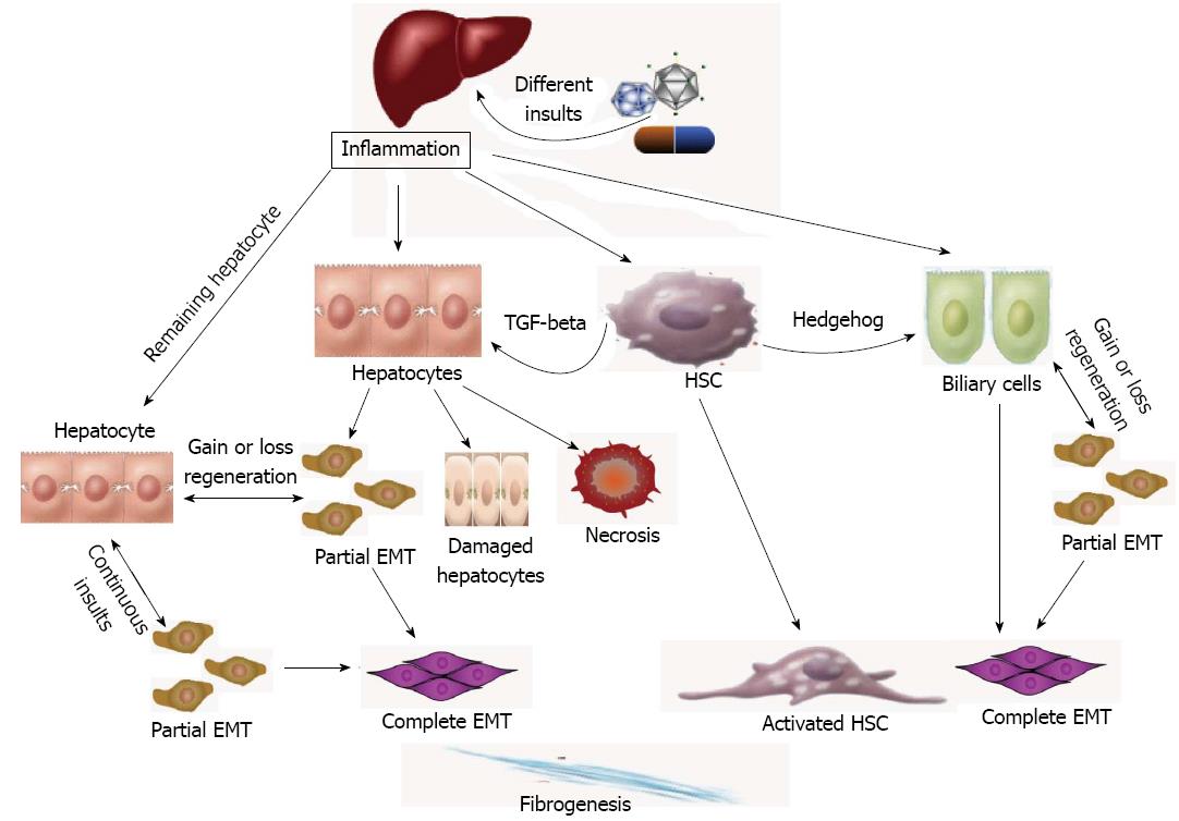
Figure 1 Liver regeneration and the epithelial to mesenchymal transition.
The diagram shows the fibrogenesis via epithelial to mesenchymal transition (EMT) in the liver. Different insults initiate inflammation and then cause hepatocyte stellate cells (HSCs) activation and hepatocyte and biliary cell damage, necrosis and EMT. Except for the necrotic and damaged cells, the remaining “normal” liver cells can be divided into 2 groups: normal and EMT-like hepatocytes. Continuous insults will shift those EMT-like cells to complete EMT cells and finally myofibroblasts, the main producer of extracellular matrix, which may be one of the main causes of an early loss of regenerative capacity. A similar process also occurs in biliary cells. HSCs play an important role in secreting key cytokines, such as transforming growth factor-beta (TGF-beta) and hedgehog, to affect the adjacent cells and promote EMTs.
- Citation: Xue ZF, Wu XM, Liu M. Hepatic regeneration and the epithelial to mesenchymal transition. World J Gastroenterol 2013; 19(9): 1380-1386
- URL: https://www.wjgnet.com/1007-9327/full/v19/i9/1380.htm
- DOI: https://dx.doi.org/10.3748/wjg.v19.i9.1380


