Copyright
©2013 Baishideng Publishing Group Co.
World J Gastroenterol. Nov 14, 2013; 19(42): 7419-7425
Published online Nov 14, 2013. doi: 10.3748/wjg.v19.i42.7419
Published online Nov 14, 2013. doi: 10.3748/wjg.v19.i42.7419
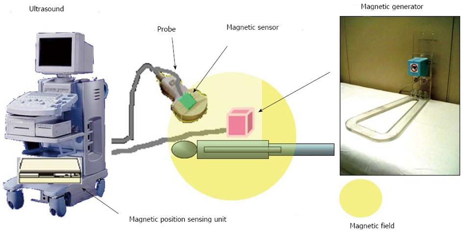
Figure 1 Real-time virtual sonography system.
Pre-scanned computed tomography volume data are processed in the main body of the ultrasound machine.
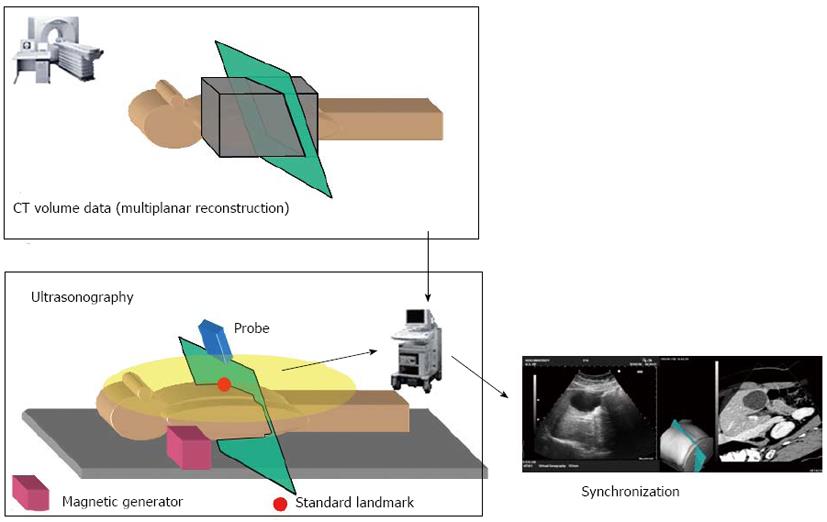
Figure 2 Calibration process.
Transfer of the obtained computed tomography (CT) volume data; configuration of the settings for the standard landmark (ensiform cartilage/aorta/portal vein/other); and positional information of the probe is sensed. According to the probe sensor, the multiplanar reconstruction image is shown as an optimal angled plane
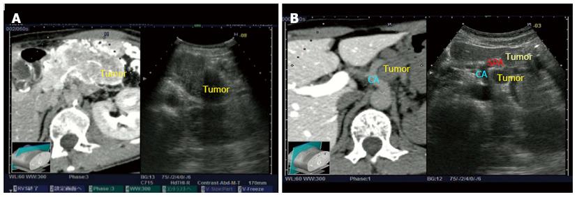
Figure 3 Pancreatic endocrine tumor (excellent case).
A: Invasive pancreatic mass (60 mm) in body tail. B: Evaluation of the relationship between celiac artery (CA) and splenic artery (SPA): there was no vascular irregularity and the boundary between the tumor and CA/SPA remained intact.
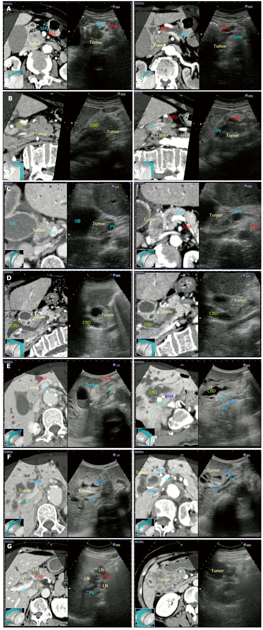
Figure 4 The resulting evaluation levels of the real-time virtual sonography images were 12 excellent (pancreatic cancer, bile duct cancer, cholecystolithiasis and cholangiocellular carcinoma) and 3 good (pancreatic cancer and gallbladder cancer).
A: Pancreatic cancer (excellent case); the positional relationships among the tumor and vessels [superior mesenteric artery (SMA) and superior mesenteric vein, (SMV)] were well evaluated; B: Pancreatic cancer (excellent case); the positional relationships among the tumor, vessels [portal vein (PV)], common bile dilatation (CBD), and main pancreatic duct (MPD) were well evaluated; C: Bile duct cancer (excellent case); the positional relationships among the tumor and vessels (SMA, SMV and PV) were well evaluated; D: Bile duct cancer (excellent case); the positional relationship between the tumor and CBD was well evaluated; E: Bile duct cancer (excellent case); the positional relationships among the tumor and vessels [proper hepatic artery (PHA), right hepatic artery (RHA), PV] were well evaluated; F: Bile duct cancer (excellent case); the positional relationships among the tumor and vessels [RHA, PV and inferior vena cava (IVC)] were well evaluated; G: Gallbladder cancer (good case); there was a gap in the positional relationships among the tumor and vessels.
- Citation: Sofuni A, Itoi T, Itokawa F, Tsuchiya T, Kurihara T, Ishii K, Tsuji S, Ikeuchi N, Tanaka R, Umeda J, Tonozuka R, Honjo M, Mukai S, Moriyasu F. Real-time virtual sonography visualization and its clinical application in biliopancreatic disease. World J Gastroenterol 2013; 19(42): 7419-7425
- URL: https://www.wjgnet.com/1007-9327/full/v19/i42/7419.htm
- DOI: https://dx.doi.org/10.3748/wjg.v19.i42.7419


