Copyright
©2013 Baishideng Publishing Group Co.
World J Gastroenterol. Jun 28, 2013; 19(24): 3895-3898
Published online Jun 28, 2013. doi: 10.3748/wjg.v19.i24.3895
Published online Jun 28, 2013. doi: 10.3748/wjg.v19.i24.3895
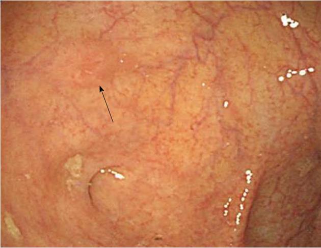
Figure 1 Initial colonoscopic findings (from another hospital).
Colonoscopic examination revealed a IIa-like, ill-defined and flatly elevated 5-mm tumor in the cecum (arrow).
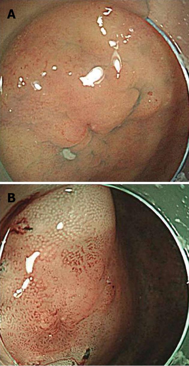
Figure 2 Colonoscopic findings at this hospital.
A: Colonoscopic examination revealed a IIa-like, ill-defined and flatly elevated 9-mm residual tumor in the cecum; B: Narrow band imaging shows the lesion more clearly.
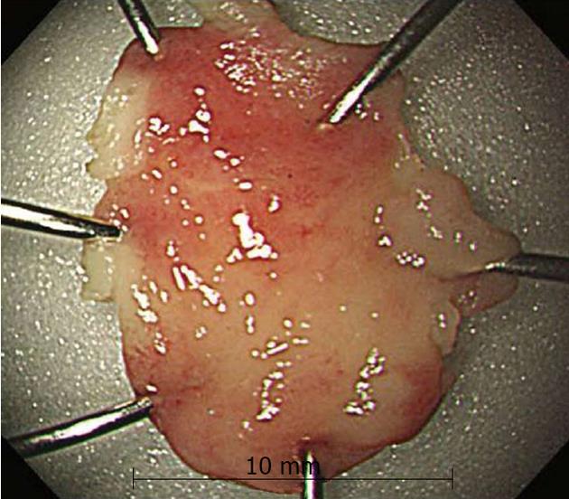
Figure 3 Resected specimen by endoscopic mucosal resection.
The resected specimen was 10 mm in diameter.
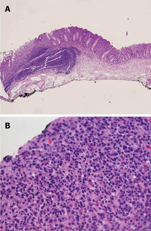
Figure 4 Scanning view of the endoscopic mucosal resection site.
Histologically, the resected specimen showed diffusely infiltrated signet ring cells in the lamina propria without venous or perineural invasion. A: hematoxylin/eosin staining, × 40; B: hematoxylin/eosin staining, × 400.
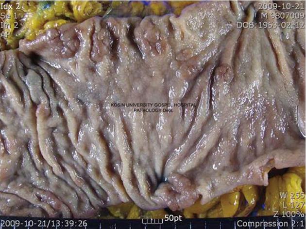
Figure 5 Gross findings.
The mucosal surface of the cecum showing an ill-defined irregularly-shaped scar (probably the endoscopic mucosal resection site). The remaining mucosa showing multiple polyps and previous polypectomy sites.
- Citation: Kim JH, Park SJ, Park MI, Moon W, Kim SE. Early-stage primary signet ring cell carcinoma of the colon. World J Gastroenterol 2013; 19(24): 3895-3898
- URL: https://www.wjgnet.com/1007-9327/full/v19/i24/3895.htm
- DOI: https://dx.doi.org/10.3748/wjg.v19.i24.3895


