Copyright
©2013 Baishideng Publishing Group Co.
World J Gastroenterol. Jan 14, 2013; 19(2): 316-318
Published online Jan 14, 2013. doi: 10.3748/wjg.v19.i2.316
Published online Jan 14, 2013. doi: 10.3748/wjg.v19.i2.316
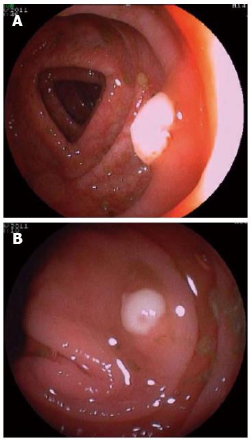
Figure 1 Colonoscopy images showing two polyps, 4 and 6 mm in size, in the right colon with adherent whitish surface.
A: Polyp of 6 mm; B: Polyp of 4 mm. The mucosa round the polyps and in the rest of colon is normal.
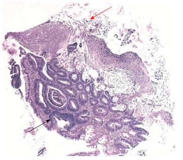
Figure 2 Low power view of a mucosal fragment.
Histopathology shows: at the bottom, tubular structures lined by columnar epithelial cells with pseudostratified nuclei, consistent with low grade tubular adenoma (black arrow); at the top, ballooned crypts and intercrypt necrosis, an exudate featuring the classical “volcano” lesion (red arrow) surrounded by a laminated pseudomembrane composed of neutrophils, mucin and fibrin (hematoxylin and eosin, x 100).
- Citation: Hernández-Rocha C, Barra-Carrasco J, Guzmán AM, Paredes-Sabja D, Lezcano G, Zoroquiaín P, Álvarez-Lobos M. Atypical presentation of pseudomembranous colitis localized in adenomatous polyps. World J Gastroenterol 2013; 19(2): 316-318
- URL: https://www.wjgnet.com/1007-9327/full/v19/i2/316.htm
- DOI: https://dx.doi.org/10.3748/wjg.v19.i2.316


